Gamma Camera
This lesson covers:
- The definition and functioning of medical tracers
- The use of technetium-99m and fluorine-18 radioactive isotopes in tracers
- The role of medical tracers in detecting various conditions
- The process of image creation using gamma cameras
Understanding medical tracers
Medical tracers are substances that emit radiation, allowing healthcare professionals to study how tissues and organs are functioning. These tracers are compounds where a radioactive isotope is attached to a molecule that the body can use, such as glucose.
Upon administration into the bloodstream, these tracers gather in regions where the body uses the tagged molecule, marking spots of higher activity or dysfunction. The decay of the radioactive isotope releases detectable radiation, which is then used to create images of the tracer distribution within the body.
Key features:
- Tracers provide insights into both the structure and function of organs, unlike traditional X-rays which only show structure.
- They concentrate in areas with high metabolic rates, indicating active processes or abnormalities.
- Specific tracers are designed to target distinct tissues and biological functions.
Utilising Technetium-99m and Fluorine-18 Isotopes
Technetium-99m and fluorine-18 are two isotopes frequently employed as tracers due to their suitable properties.
Technetium-99m
- Known for emitting gamma radiation, which gamma cameras can detect.
- Its half-life of 6 hours strikes a balance, offering ample time for imaging without prolonged radioactive exposure.
Fluorine-18
- It decays by positron emission, making it ideal for PET (Positron Emission Tomography) scans.
- A relatively short half-life of 110 minutes helps in reducing the radiation dose to the patient.
These isotopes are bound to substances like glucose or water, creating tracers that accumulate in the body's areas where these substances are naturally used.
Applications of medical tracers
Medical tracers are instrumental in diagnosing and studying various conditions:
Damaged Heart Tissue
- Areas with reduced blood flow can indicate heart tissue damage or blockages in the coronary artery.
- Tracers help identify heart attacks by highlighting areas of dead muscle tissue.
Cancer Imaging
- Due to their high metabolic rate, cancer cells consume more glucose, making glucose-based tracers ideal for identifying tumours.
Brain Imaging
- Tracers are used to map cerebral blood flow and metabolic activity, valuable for diagnosing disorders like Alzheimer’s and Parkinson’s disease.
Imaging with gamma cameras
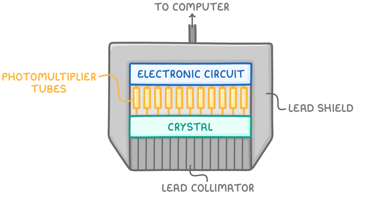
Gamma cameras are sophisticated devices positioned outside the body to capture the radiation emitted by tracers. They consist of:
- Lead shielding to prevent background radiation from interfering.
- A collimator that only permits gamma rays entering at right angles.
- A crystal that lights up (scintillates) upon gamma ray impact.
- Photomultiplier tubes that convert the scintillations into electrical signals.
- A computer system that translates these signals into images, indicating the tracer distribution.
Through these images, doctors can assess organ function and detect abnormalities without invasive procedures, enhancing diagnostic accuracy and patient care.