X-Rays
This lesson covers:
- The components and functioning of an X-ray tube
- How X-rays are produced when electrons strike the tungsten anode
- Calculating the energy and wavelength of emitted X-rays
- Increasing the intensity of the X-ray beam
- The attenuation of X-rays as they pass through matter
- The greater absorption of X-rays by bone compared to soft tissue
- The use of X-rays in CAT scans
X-ray tube components and functioning
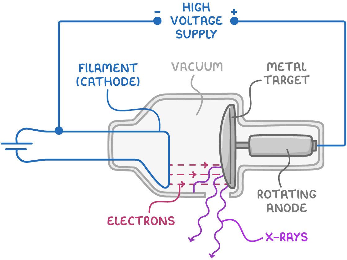
An X-ray tube operates as a sophisticated electrical device comprising:
- Cathode: This component emits a focused beam of electrons when heated by an electric current passing through a filament. It's typically shaped like a cup to direct the electrons efficiently.
- Anode: A target metal, often tungsten, where the electron beam is aimed. It is propelled by a high voltage across the tube, which accelerates the electrons towards it.
Upon striking the tungsten anode, the interaction between the electrons and the tungsten not only generates X-rays but also significant heat. To manage this heat, the anode is designed to rotate swiftly, allowing a copper mount to disperse the heat effectively.
Worked example - Calculating maximum X-ray energy
X-rays are produced in an X-ray tube with an accelerating potential of 100 kV. Calculate the maximum X-ray energy.
Step 1: Convert kV to V
to convert from kV to V, multiply by 1,000
100 kV = 100,000 V
Step 2: Formula
E = e V
Step 3: Substitution and correct evaluation
E = 1.6 x 10-19 x 100,000 = 1.6 x 10⁻¹⁴ J
Calculating X-ray wavelength from energy
For photons, including X-rays:
E = λh c
Where:
- E = photon energy (J)
- h = Planck’s constant ( 6.63 x 10⁻³⁴ J s)
- c = speed of light (3 x 10⁸ m s-1)
- λ = wavelength (m)
This equation implies that halving the maximum energy by reducing the potential difference will result in doubling the wavelength of the emitted X-rays.
Increasing X-Ray beam intensity
To enhance the X-ray beam's intensity, one can:
- Increase the accelerating potential difference, which heightens the energy of the electrons.
- Boost the heating current of the filament to produce more electrons per second.
These actions elevate the number of X-ray photons emitted, thereby increasing the beam's intensity.
Attenuation of X-Rays
X-rays diminish in intensity, or attenuate, as they traverse matter, primarily due to:
- Absorption
- Scattering
This attenuation follows an exponential decrease with depth, as described by:
I=I0e−μx
Where:
- I = intensity after traversing a distance x (W m-2)
- I0 = initial intensity (W m-2)
- μ = attenuation coefficient (cm-1)
- x = depth within the material (m)
Worked example - Calculating attenuation of X-rays
Calculate the X-ray intensity after passing through 5 cm of material with an initial intensity of 1,000 W m-2. The attenuation coefficient of the material is 0.2 cm-1.
Step 1: Formula
I=I0e−μx
Step 2: Substitution and correct evaluation
I = 1,000 e−0.2×5 = 367.9 W m−2
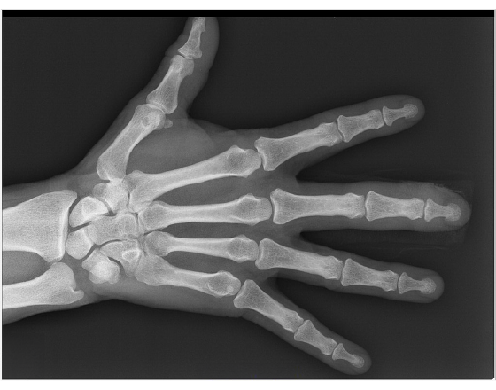
Contrast between bone and soft tissue
Due to their higher atomic numbers, bones absorb X-rays more efficiently than soft tissues, making them more prominent on X-ray images. Contrast can be further improved by administering contrast media, such as barium or iodine, which are visible on X-rays and help in visualising internal structures.
X-Rays in CAT Scans
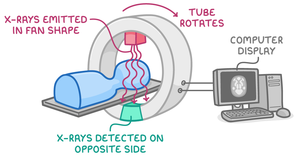
Computerised Axial Tomography (CAT), or CT scans, use:
- A rotating X-ray beam and detectors to capture images of body slices.
- These images are processed to produce detailed cross-sectional views of soft tissues.
- Combining multiple slices generates three-dimensional models of internal body structures.