Non-specific Animal Defences: Phagocytosis
This lesson covers:
- The role of phagocytes in the immune response
- The process of phagocytosis
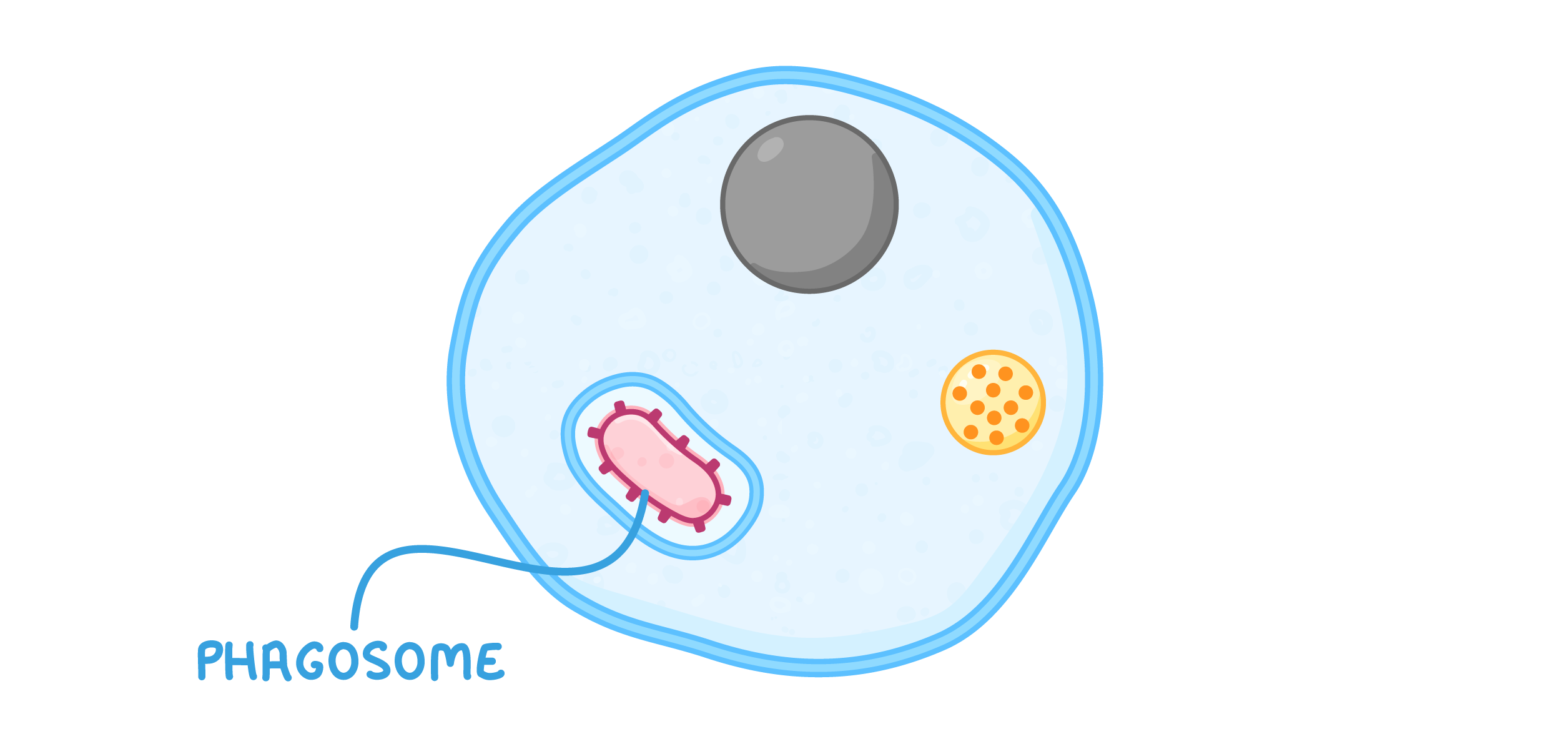
Phagocytes Phagocytosis is a type of non-specific defence involving a type of cell known as a phagocyte. 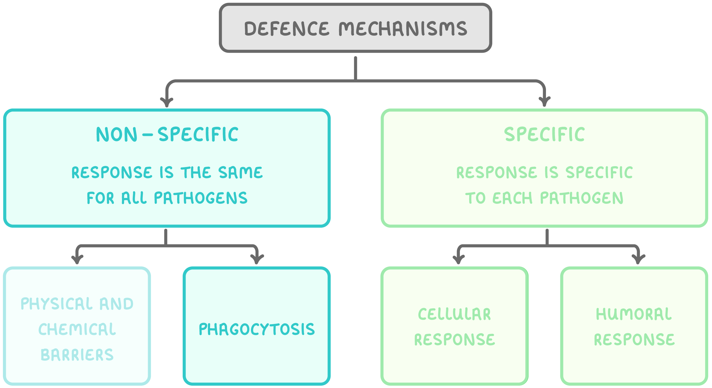 |
Phagocytes are a type of white blood cell that engulf and destroy pathogens (phagocytosis). They are found in the blood and body tissues of many organisms. There are two main types of phagocyte:
|
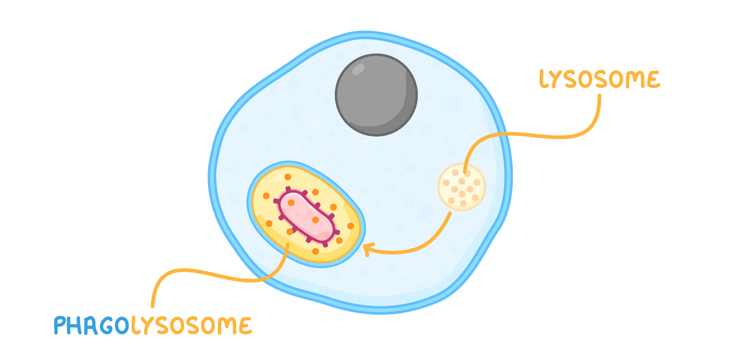
Phagocytosis The process of phagocytosis is carried out as follows:  The pathogen releases chemicals that attract a phagocyte. The diagrams above only show one phagocyte and one pathogen, but in reality many phagocytes and many pathogens interact at the same time. |


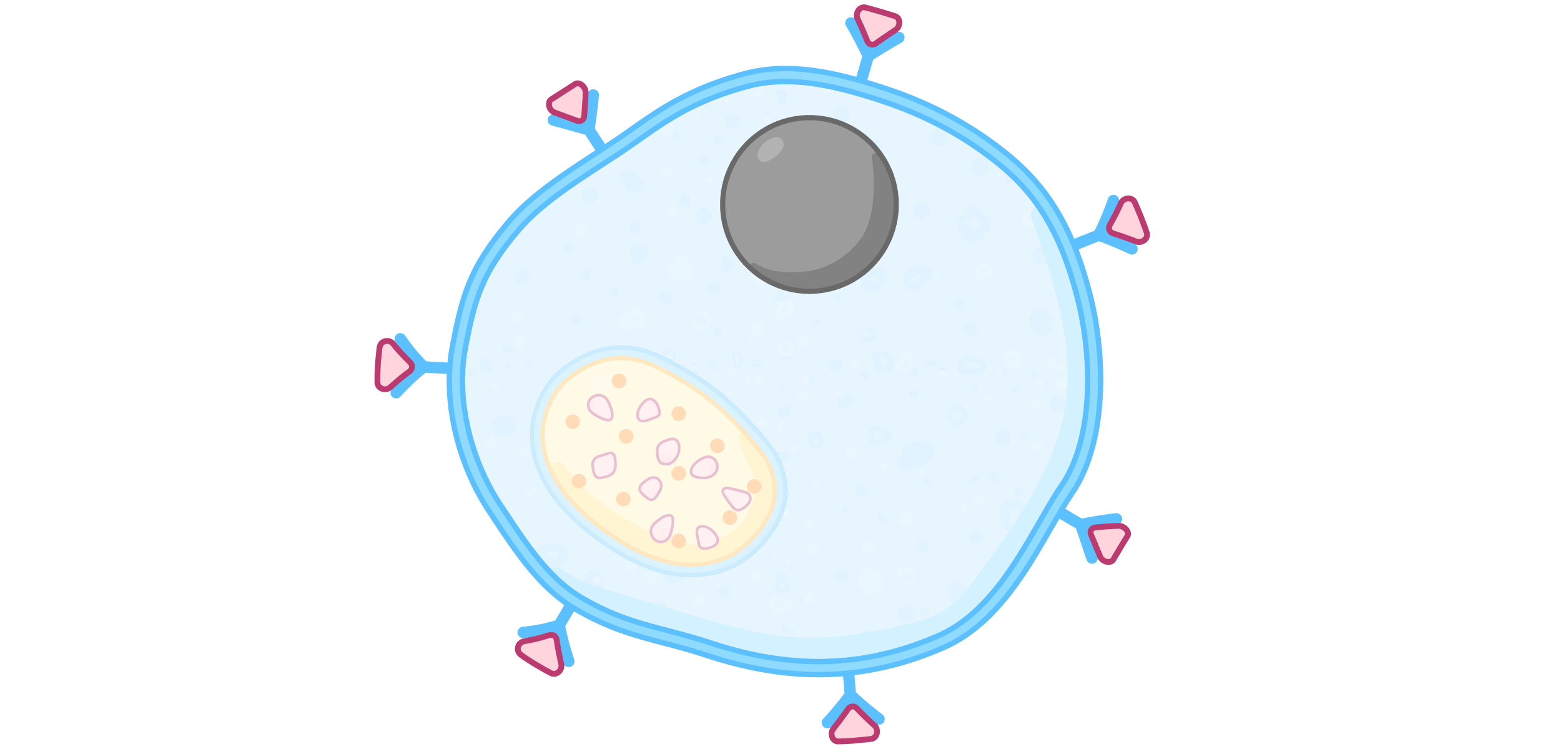

Cytokines and opsonins Cytokines and opsonins are molecules that help with the process of phagocytosis. Cytokines are chemicals released by phagocytes that have engulfed a pathogen. They act as cell-signalling molecules to trigger the movement of other phagocytes to the site of infection. Cytokines also trigger an increase in body temperature which inhibits the reproduction of pathogens and allows the specific immune system to work faster. Opsonins are chemicals (e.g. antibodies) that bind to pathogens to make them easily recognisable by phagocytes. Phagocytes contain receptors on their cell-surface which bind to common opsonins, making it easier for the phagocyte to bind to the pathogen and destroy it. |