Structure of Skeletal Muscle
This lesson covers:
- The structure of skeletal muscle fibres
- The components within muscle fibres that allow contraction
The structure of skeletal muscle fibres
Skeletal muscles comprise numerous bundles of long, cylindrical muscle fibres.
Individual cells are fused to form muscle fibres during development to avoid weakness at the junctions between cells and to increase overall muscle strength.

Key components of a muscle fibre include:
- Sarcolemma - The cell-surface membrane of a muscle fibre.
- Sarcoplasm - The cytoplasm of a muscle fibre.
- Transverse tubules (T tubules) - Extensions of the sarcolemma that transmit electrical signals, to ensure that the entire muscle receives the impulse to contract simultaneously.
- Sarcoplasmic reticulum - A specialised endoplasmic reticulum that is responsible for storing and releasing calcium ions.
- Myofibrils - Subcellular structures designed for contraction.
- Multiple nuclei - Skeletal muscle fibres have many nuclei because several cells merge to form one muscle fibre.
- Mitochondria - These release energy in the form of ATP for muscle contraction.
The structure of myofibrils
Myofibrils, the core units of muscle fibres, contain organised bundles of protein filaments.
These filaments slide past each other to enable muscle contraction.
The structure of a sarcomere
Myofibrils are made up of repeating units called sarcomeres.
Main filaments in a sarcomere:
- Myosin filaments - Thick filaments composed of long rod-shapes with bulbous heads that project to the side.
- Actin filaments - Thin filaments composed of two strands twisted around each other.
Under a microscope, myofibrils exhibit a distinctive banding pattern, which corresponds to the arrangement of myosin and actin within each sarcomere.
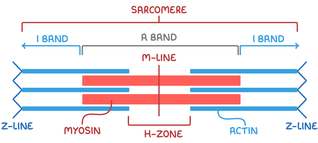
Key sections of the sarcomere:
- A band (dark band) - Area with both myosin and overlapping actin filaments (darker in colour).
- I band (light band) - Area containing only light actin filaments (lighter in colour).
- Z-lines - Mark the boundaries of each sarcomere unit (central line within the I band).
- M-line - The central line of a sarcomere.
- H-zone - Area with only myosin filaments (central lighter region in the A band).