Eukaryotic Cells: Animal Cells

This lesson covers:

  1. The difference between eukaryotes and prokaryotes
  2. The structure of animal cells 
  3. The functions of their cell parts

Eukaryotes and Prokaryotes

Living organisms can be divided into two main groups:

  1. Eukaryotes - These are (mostly) multicellular organisms made up of eukaryotic cells like animals, plants, fungi, and protists.
  2. Prokaryotes - These are single-celled organisms made up of prokaryotic cells like bacteria.


Eukaryotic cells are more complex, contain membrane-bound organelles, and have their DNA in the form of chromosomes within a nucleus. 

Animal cells

The diagram below shows all the organelles found in a typical animal cell. 

Diagram showing the organelles in a typical animal cell including nucleus, nucleolus, rough endoplasmic reticulum, smooth endoplasmic reticulum, mitochondrion, lysosome, ribosomes, and Golgi apparatus.

Nucleus

The nucleus is the largest organelle within an animal cell.

Diagram of an animal cell nucleus showing nuclear pore, nucleolus, chromatin, and nuclear envelope.

Structure: 

  • Contains genetic information in the form of chromosomes.
  • Contains a structure known as a nucleolus.
  • Surrounded by a nuclear envelope (double membrane) containing pores.


Functions: 

  • Controlling the cell's activities - The DNA contains instructions to produce proteins. 
  • Synthesis of ribosomes - The nucleolus makes ribosomal RNA.
  • Exchange between nucleus and cytoplasm - Substances can enter or leave the nucleus via the nuclear pores.

Cell-surface membrane

The cell-surface membrane is also known as the plasma membrane. 
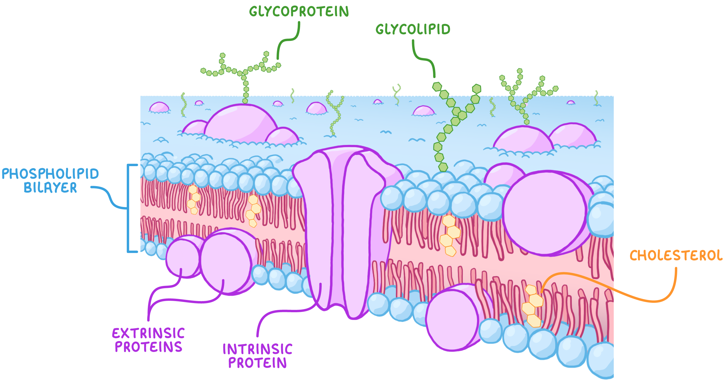
Illustration of the cell-surface membrane structure showing phospholipid bilayer, extrinsic proteins, intrinsic proteins, glycoprotein, glycolipid, and cholesterol.

Structure: 

  • Found on the surface of animal cells.
  • Mainly made up of lipids and protein.


Functions: 

  • Controls movement of substances into and out of the cell - The membrane is partially permeable.
  • Cell signalling - Receptors can detect signals from other cells.


We cover the structure in more detail in our lesson on the phospholipid bilayer.

Mitochondria

Mitochondria are found in large numbers in cells that requires lots of energy.
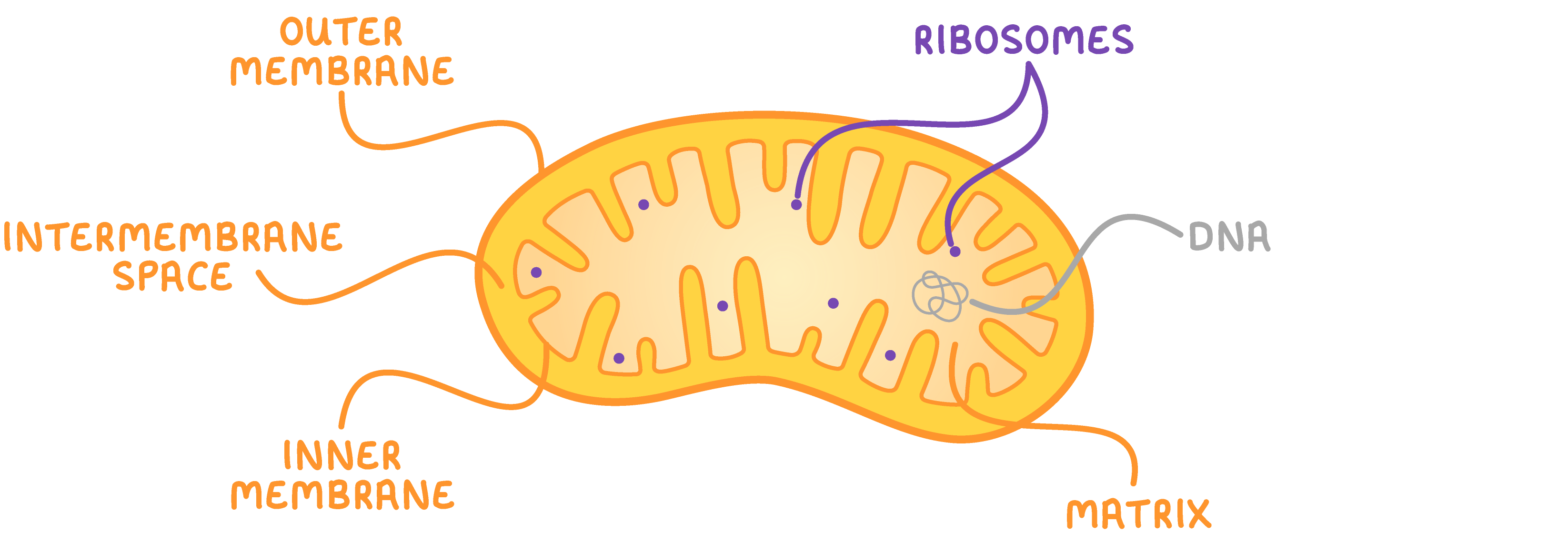
Diagram showing the structure of a mitochondrion including outer membrane, intermembrane space, inner membrane, matrix, ribosomes, and DNA.

Structure: 

  • Contain an enzyme-rich liquid known as the matrix. 
  • Surrounded by a double membrane in which the inner membrane is folded to form structures known as cristae.
  • Contain their own DNA (mtDNA) and ribosomes.


Function: 

  • Site of aerobic respiration - Produces ATP.

Ribosomes

Ribosomes are very small organelles found in the cytoplasm or attached to rough endoplasmic reticulum. 


The size of ribosomes are measured in S units. Eukaryotic cells contain 80S ribosomes.

Illustration showing the structure of a ribosome with large and small subunits.

Structure: 

  • Made up of proteins and rRNA (ribosomal RNA).
  • Consist of a large and a small subunit.
  • Not surrounded by a membrane. 


Function: 

  • Site of protein synthesis - Involved in the process of translation.

Golgi apparatus

The Golgi apparatus, sometimes called the Golgi body, consists of cisternae and vesicles.
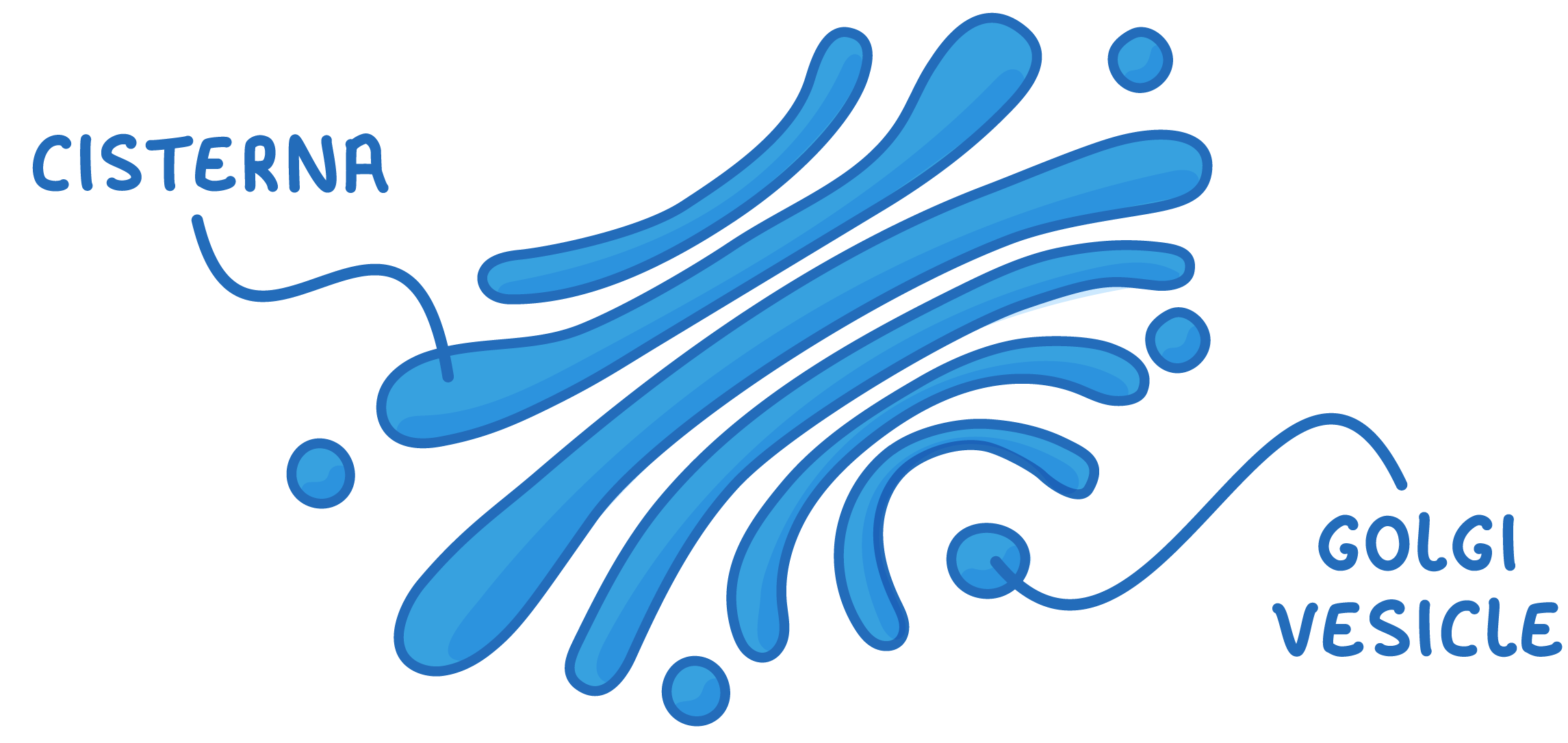
Diagram showing the structure of the Golgi apparatus with cisterna and Golgi vesicle labeled.

Structure: 

  • Contain fluid-filled, membrane-bound sacs known as cisternae.
  • Contain smaller vesicles.


Functions: 

  • Process and package lipids and proteins - Carried out by the cisternae.
  • Store and transport lipids and proteins - Carried out by the vesicles.
  • Synthesise lysosomes - Specialised vesicles. 

Rough endoplasmic reticulum (RER)

Rough endoplasmic reticulum is covered with ribosomes (that's why it's rough - the ribosomes cause the little bumps). 
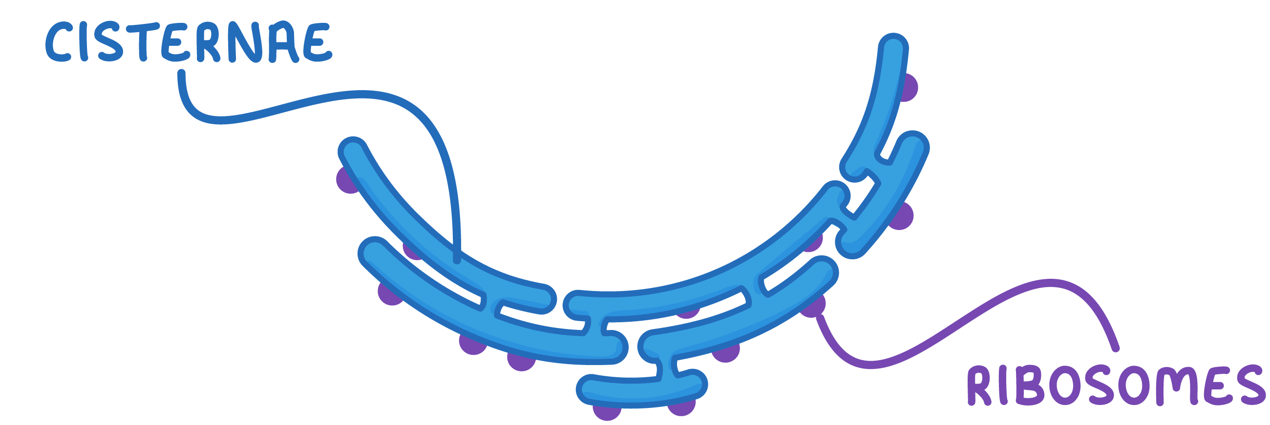
Diagram showing the structure of rough endoplasmic reticulum with cisternae and ribosomes.

Structure: 

  • Contains a network of membranes enclosing a fluid-filled space, known as cisternae. 
  • The surface of the cisternae is covered with ribosomes. 


Function: 

  • Synthesis and transport of proteins - The proteins are made using the ribosomes.

Smooth endoplasmic reticulum (SER)

Smooth endoplasmic reticulum lacks ribosomes. 

Diagram showing the structure of cisternae in the endoplasmic reticulum.

Structure: 

  • Same as the rough endoplasmic reticulum without the ribosomes.


Function: 

  • Synthesis, storage, and transport of lipids and carbohydrates - For example, cholesterol and steroid hormones.

Lysosomes

Lysosomes are round organelles with no clear internal structure.
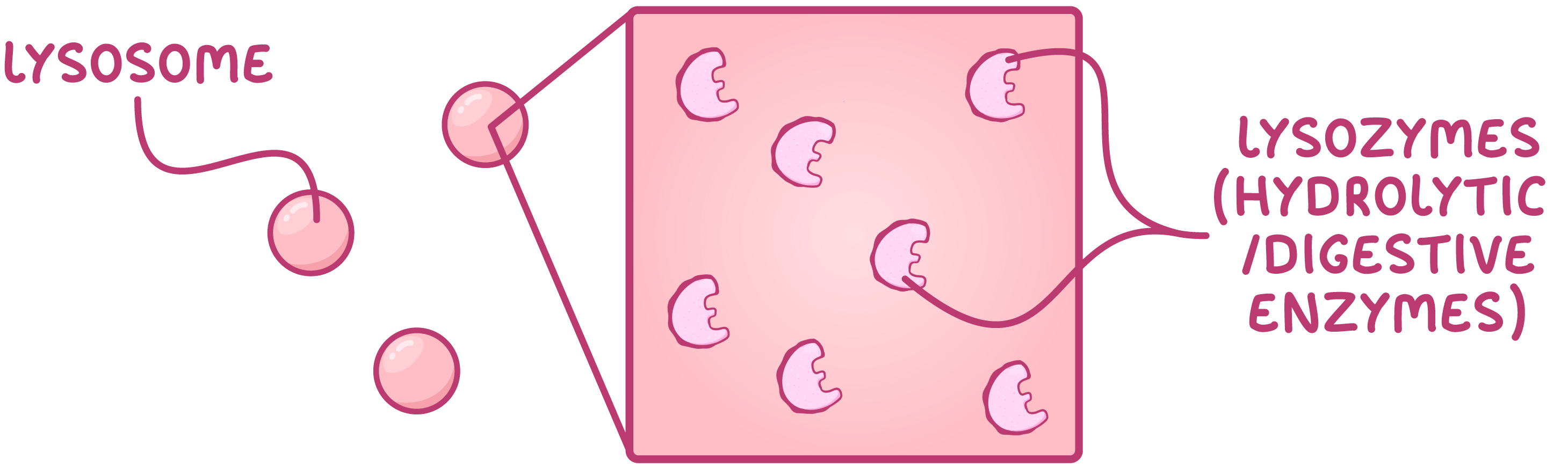
Diagram showing the structure of a lysosome and the presence of hydrolytic digestive enzymes.

Structure: 

  • Contain hydrolytic enzymes. 
  • Surrounded by a membrane to keep enzymes separate from the cytoplasm of the cell.


Functions: 

  • Digest invading cells - This process uses enzymes. 
  • Break down waste material - This material includes old organelles. 

Cytoskeleton

The cytoskeleton is present throughout the cytoplasm and provides structure and support to the cell. It consists of 3 main components: microfilaments, microtubules, and intermediate filaments.

Microfilaments:

  • Made of the protein actin.
  • Involved in cell movement and locomotion, such as crawling and muscle contraction.

Microtubules:

  • Made of the protein tubulin.
  • Form a scaffold-like structure throughout the cell.
  • Form the main component of the mitotic spindle, used for cell division.
  • Act as tracks for intracellular transport of vesicles and organelles.

Intermediate filaments:

  • Maintain the position of organelles within the cell.
  • Responsible for the mechanical strength of certain tissues, such as the skin and hair.