Sensory Receptors
This lesson covers:
- How receptor cells detect specific stimuli
- Examples of different types of receptor cell
- How receptor cells work
- The structure and function of Pacinian corpuscles
What is a sensory receptor?
Sensory receptors are specialised cells that detect stimuli from the environment.
Receptors act as transducers. A transducer converts one form of energy into another. In a receptor, a transducer converts the stimulus energy into a nerve impulse that is passed to the central nervous system.
Types of receptor cell
Receptor cells are designed to detect only one particular type of stimulus. The body contains a range of receptors, each tailored to respond to specific stimuli.
| Receptor type | Stimulus detected | Location |
|---|---|---|
| Photoreceptors | Light | Eyes |
| Chemoreceptors | Chemicals | Nose, tongue, blood vessels |
| Mechanoreceptors | Pressure, movement | Skin, muscles, inner ear |
| Thermoreceptors | Temperature | Skin |
How receptor cells work
There are several stages involved in how receptor cells function from detecting a stimulus to generating a nerve impulse in a neurone.
Stages of receptor cell function:
- At rest, the receptor cell-surface membrane has a voltage across it due to differences in ion concentration inside and outside the cell, known as the resting potential.
- When a stimulus is detected, the cell-surface membrane becomes more permeable, allowing more ions to flow in and out.
- This alters the membrane's voltage, creating what is called the generator potential (or receptor potential).
- A larger stimulus results in a bigger change in voltage, producing a larger generator potential.
- If the generator potential reaches a threshold level, it triggers an action potential, which is an electrical signal sent along a neurone.
More action potentials indicate a stronger stimulus. If the stimulus is too weak, the threshold isn't reached and no action potential is generated.
Structure and function of Pacinian corpuscles
Pacinian corpuscles are mechanoreceptors in the skin that detect pressure and vibrations.
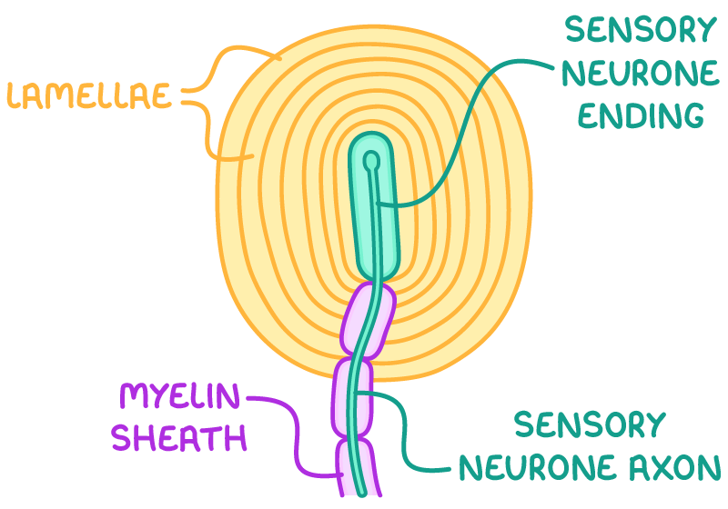
They contain the ending of a sensory neurone wrapped in layers of connective tissue called lamellae. The Pacinian corpuscle's lamellae allow it to detect pressure and vibration, demonstrating how receptors only respond to specific stimuli. There is viscous gel between each layer of tissue.
When the Pacinian corpuscle is stimulated:
- The lamellae deform, pressing on the sensory neurone ending.
- This stretches the neurone's membrane, causing it to change shape.
- This opens stretch-mediated sodium ion (Na+) channels in the membrane, increasing its permeability to Na+.
- Na+ diffuses into the neurone, depolarising it and resulting in a generator potential.
- If this signal reaches the threshold, an action potential is triggered.