Magnification and Resolution
This lesson covers:
- The definitions of magnification and resolution
- How to calculate magnification
- How to calibrate a microscope
Magnification Magnification is how many times larger an image is than the object. For example, here the image is 1,000 larger than the object, so its magnification is x 1,000. 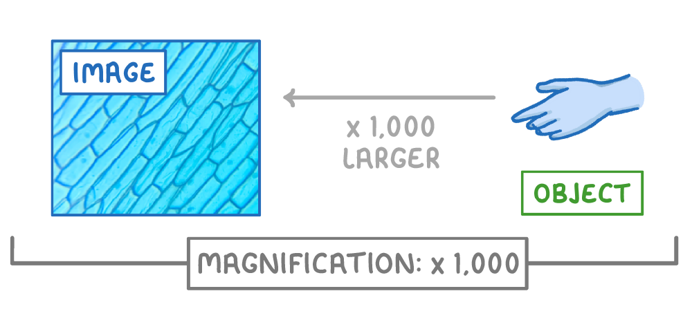 |
Resolution Resolution is the ability to distinguish between two separate points (or how detailed the image is). For example, both images here have the same magnification, but the left image has a higher resolution. 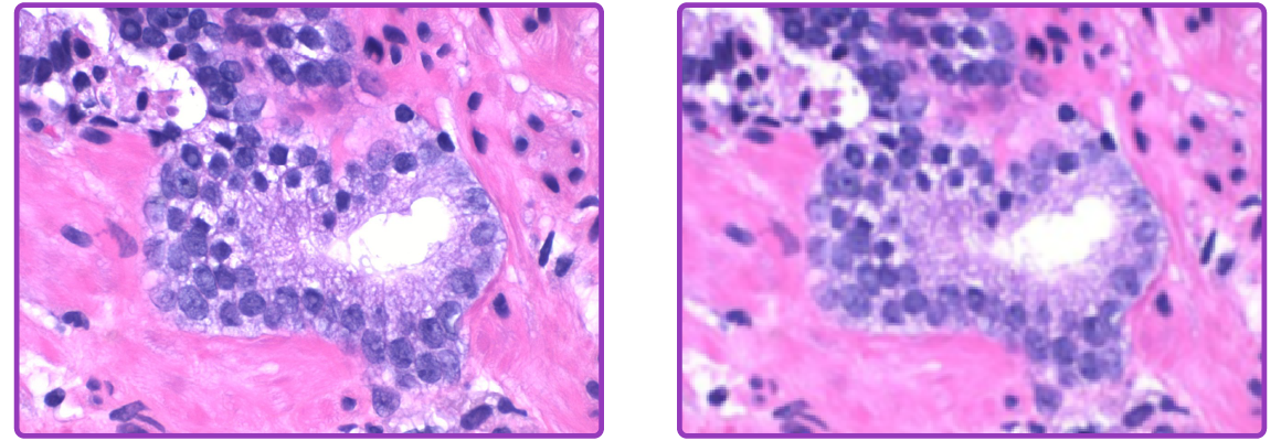 |
Calculating magnification To calculate magnification, you need to know the image size and the object size:
The equation for calculating magnification is: |
When calculating magnification, all lengths must be in the same unit. This may require converting from one unit to another. 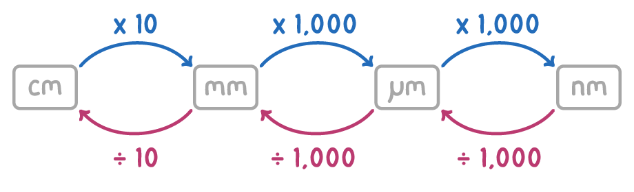 |
Calibrating a microscope When viewing a specimen, you can measure its length or width using an eyepiece graticule. This is a small scale (usually from 1 to 100) placed within the eyepiece. |
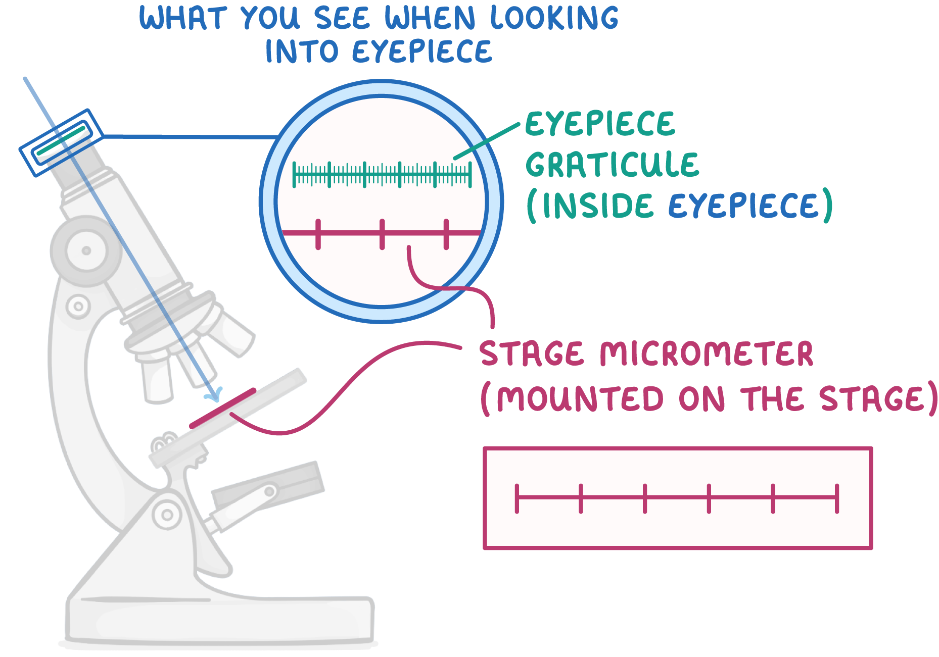 |
However, the scale divisions of the eyepiece graticule will represent different real-world distances, depending on the magnification of the objective lens you are looking through. This means the graticule needs to be calibrated for each objective lens. A stage micrometer is used to calibrate the eyepiece graticule. This is a glass slide with a scale measured in µm. The graticule is calibrated as follows:
|
Worked example - Calibrating a light microscope 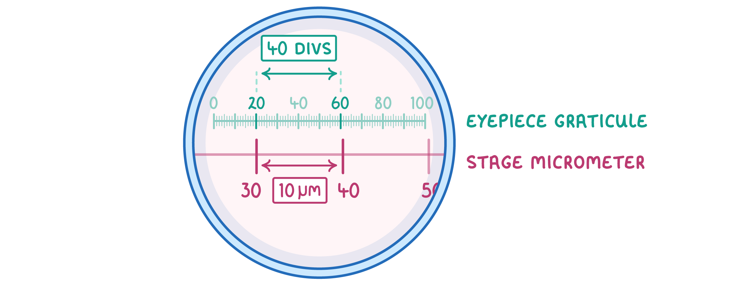 The image above represents what you'd see if you looked into the eyepiece of a light microscope. It shows a stage micrometer lined up with a graticule scale. Each division on the micrometer represents 10 µm. Calculate the size of each graticule division. |
Step 1: Count the number of graticule divisions in one micrometer division 40 graticule divisions = 1 micrometer division Step 2: Equation Step 3: Substitution and correct evaluation µm |
Worked example - Calculating magnification
A plant cell in a micrograph measures 40 mm in width. If the real width of the plant cell is actually 20 µm, what is the magnification?
Step 1: Equation
magnification =object sizeimage size
Step 2: Conversion
to convert from mm into μm, multiply by 1,000
image size =40×1,000=40,000 μm
object size =20 μm
Note: the units for image size and object size need to be the same before they are put into the magnification calculation
Step 3: Substitution and correct evaluation
magnification =2040,000
magnification = x20,000
Worked example - Calculating image size
A sperm cell is 5 µm long. How long will it appear in mm when viewed under a microscope with a magnifying power of x 1,500?
Step 1: Equation
magnification =object sizeimage size
Step 2: Rearrangement
image size = magnification× object size
Note: we need to rearrange the equation to make image size the subject
Step 3: Substitution and correct evaluation
image size =1,500×5
image size =7,500 µm
Step 4: Conversion
to convert µm into mm, divide by 1,000
image size =1,0007,500
image size =7.5 mm