Specialised Cells

This lesson covers:

  1. What 'specialised' cells are 
  2. Examples of how cells are specialised for their functions 

Specialised cells 

Like we saw in the previous lesson, all cells start out as unspecialised stem cells. These stem cells can then differentiate to form specialised cells.

Diagram showing stem cell differentiation into red blood cell, sperm cell, and white blood cell.

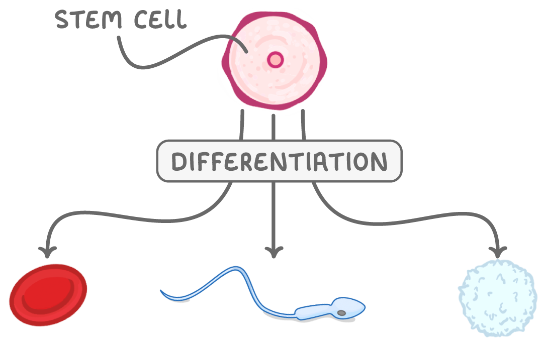
Specialised cells have features that enable them to carry out their specific function. For example, sperm cells have a tail that helps them to swim to fertilise an egg cell. 

Specialised animal cells

You need to be able to explain how the following animal cells are specialised for their functions. 

Illustration of a red blood cell showing its biconcave shape.

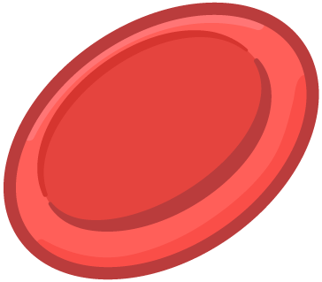
Erythrocytes

Erythrocytes or red blood cells are responsible for transporting oxygen around the body. 


Specialised features:

  • Flattened biconcave shape - This increases the surface area to volume ratio to allow diffusion of oxygen. 
  • No nucleus or organelles - This provides more room for haemoglobin (the molecule that binds to oxygen). 
  • Flexible - This is so they can fit through narrow capillaries. 
Illustration of an animal cell showing the cell membrane, nucleus, and lysosomes.

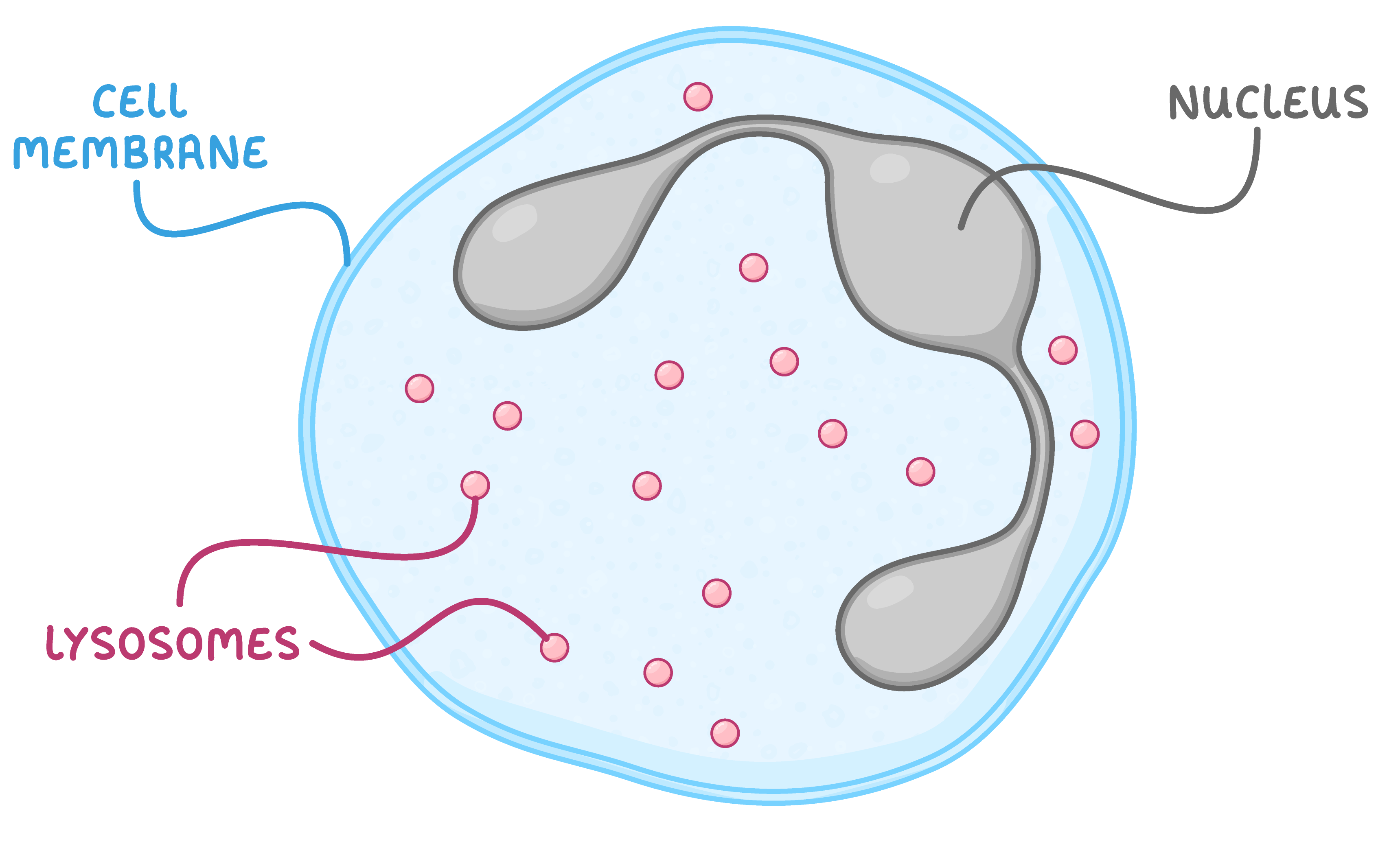
Neutrophils 

Neutrophils are types of white blood cell that help to defend the body against pathogens. 


Specialised features: 

  • Flexible cell membrane - This allows the cell to engulf pathogens. 
  • Contain many lysosomes - These contain digestive enzymes to break down engulfed particles.
  • Multi-lobed nucleus - This allows cells to deform so they can squeeze through small gaps to reach sites of infection. 
Diagram of a sperm cell showing the acrosome, mitochondria, and flagellum.

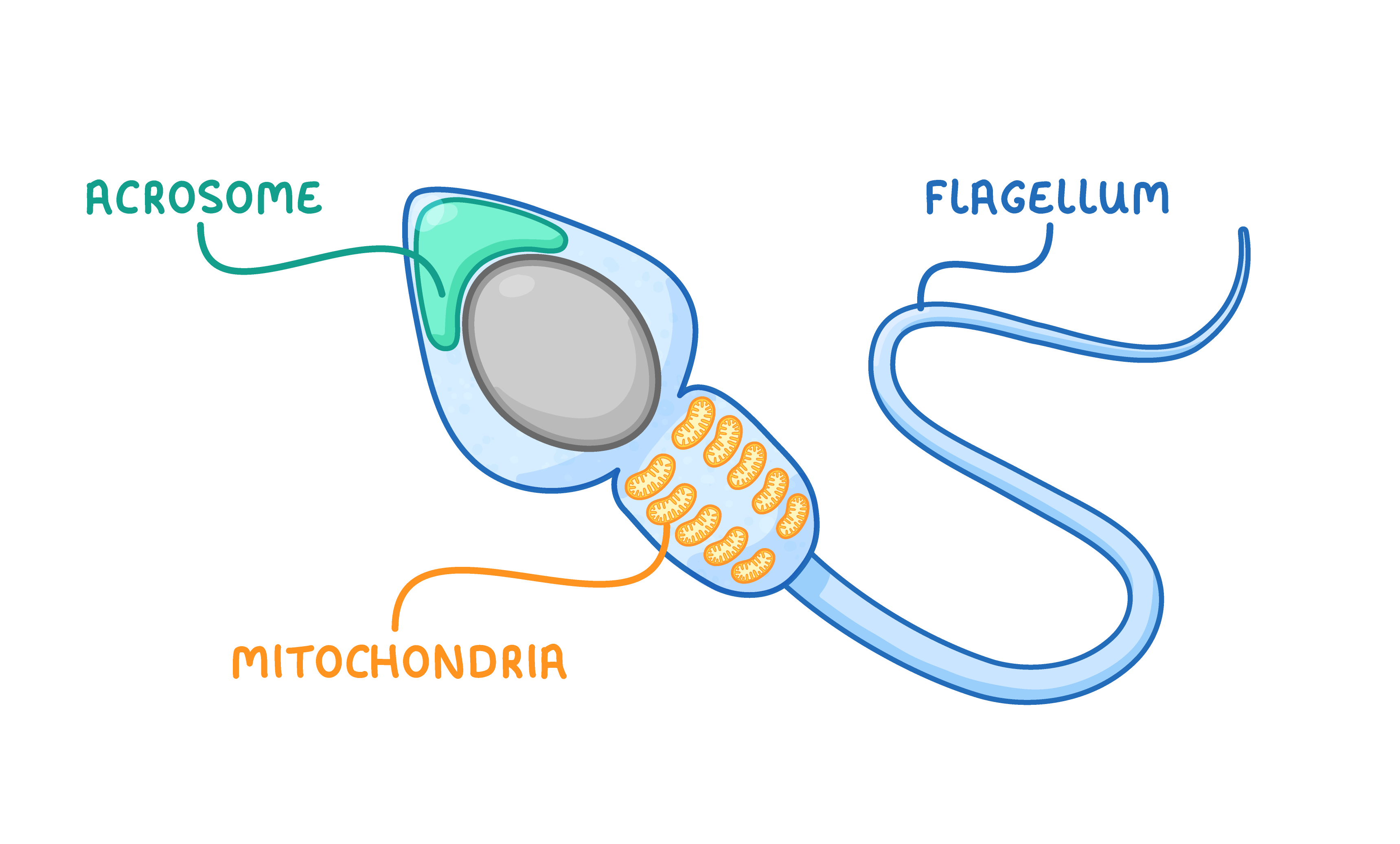
Sperm cells

Sperm cells (male gametes) carry genetic information to the female gamete.


Specialised features: 

  • Flagellum (tail) - This allows the cell to swim to the egg cell. 
  • Many mitochondria - These supply the energy needed for movement. 
  • Acrosome containing digestive enzymes - These digest the protective layers around the egg cell to allow the sperm cell to enter it.
Diagram showing specialised animal cells with a focus on their structure and function.

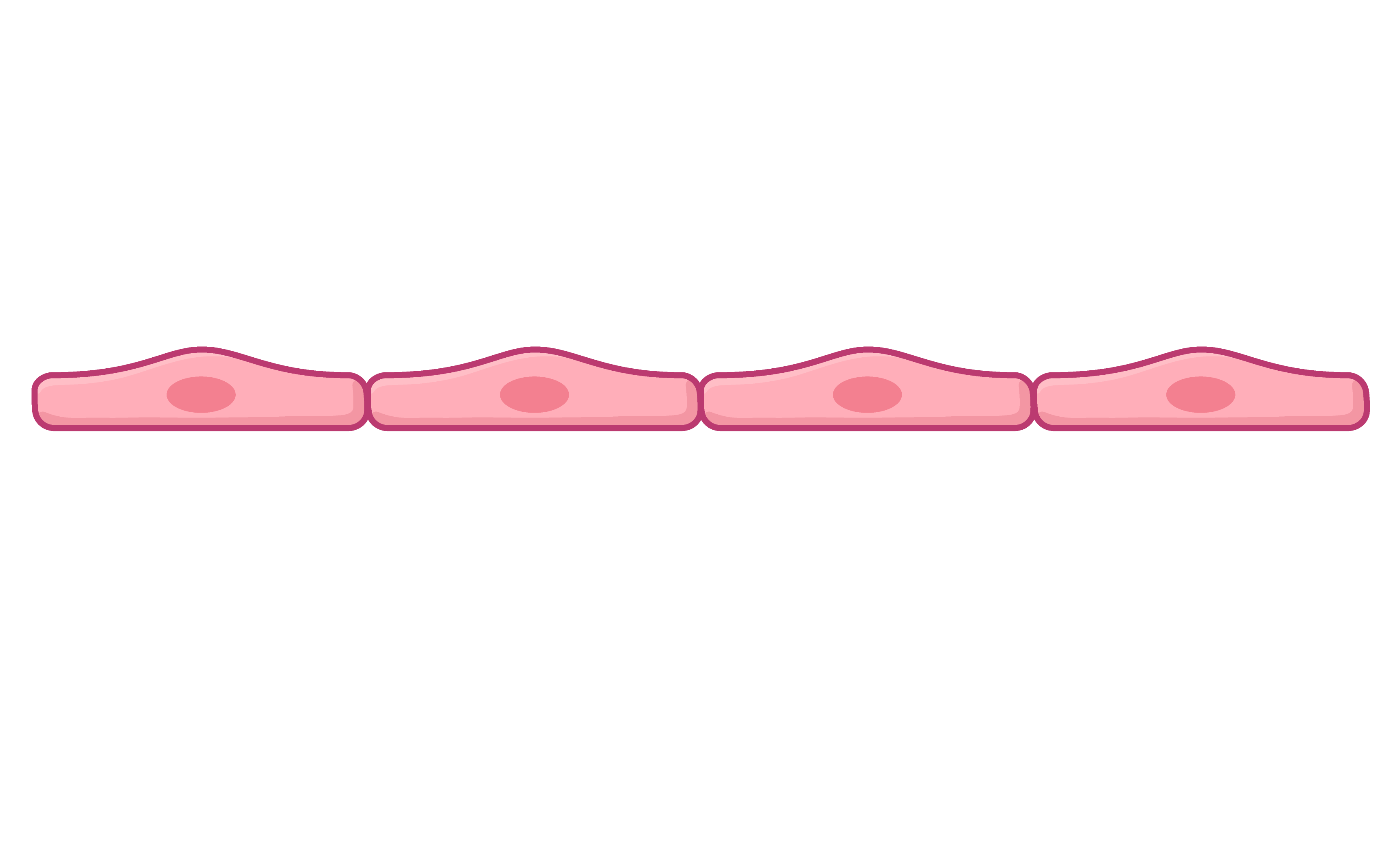
Squamous epithelial cells

Squamous epithelial cells cover the surface of organs such as the lungs and blood vessels. 


Specialised features: 

  • Very thin - This allows efficient diffusion of gases such as oxygen and carbon dioxide. 
  • Permeable - This allows diffusion of gases. 
Illustration of a ciliated animal cell showing cilia on the surface.

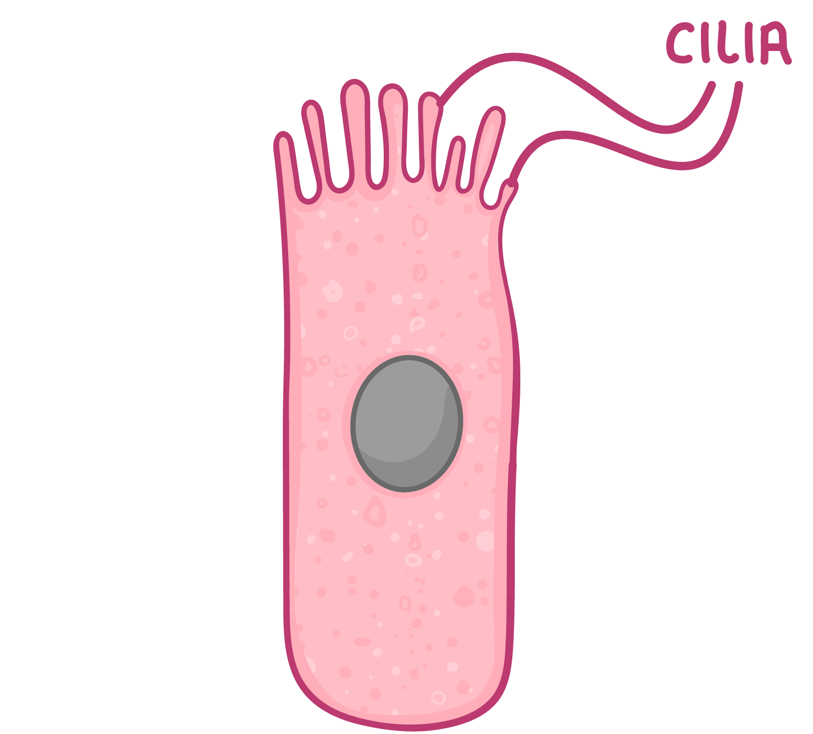
Ciliated epithelial cells

Ciliated epithelial cells also cover the surface of organs where they can move substances such as mucus or egg cells. They are found in organs such as the bronchioles and fallopian tubes. 


Specialised features: 

  • Cilia (hair-like structures) - These beat to move pathogens and mucus away from the lung or egg cells towards the uterus. 

Specialised plant cells 

You need to be able to explain how the following plant cells are specialised for their functions.

Diagram of a plant cell showing nucleus, chloroplast, cell wall, and vacuole.

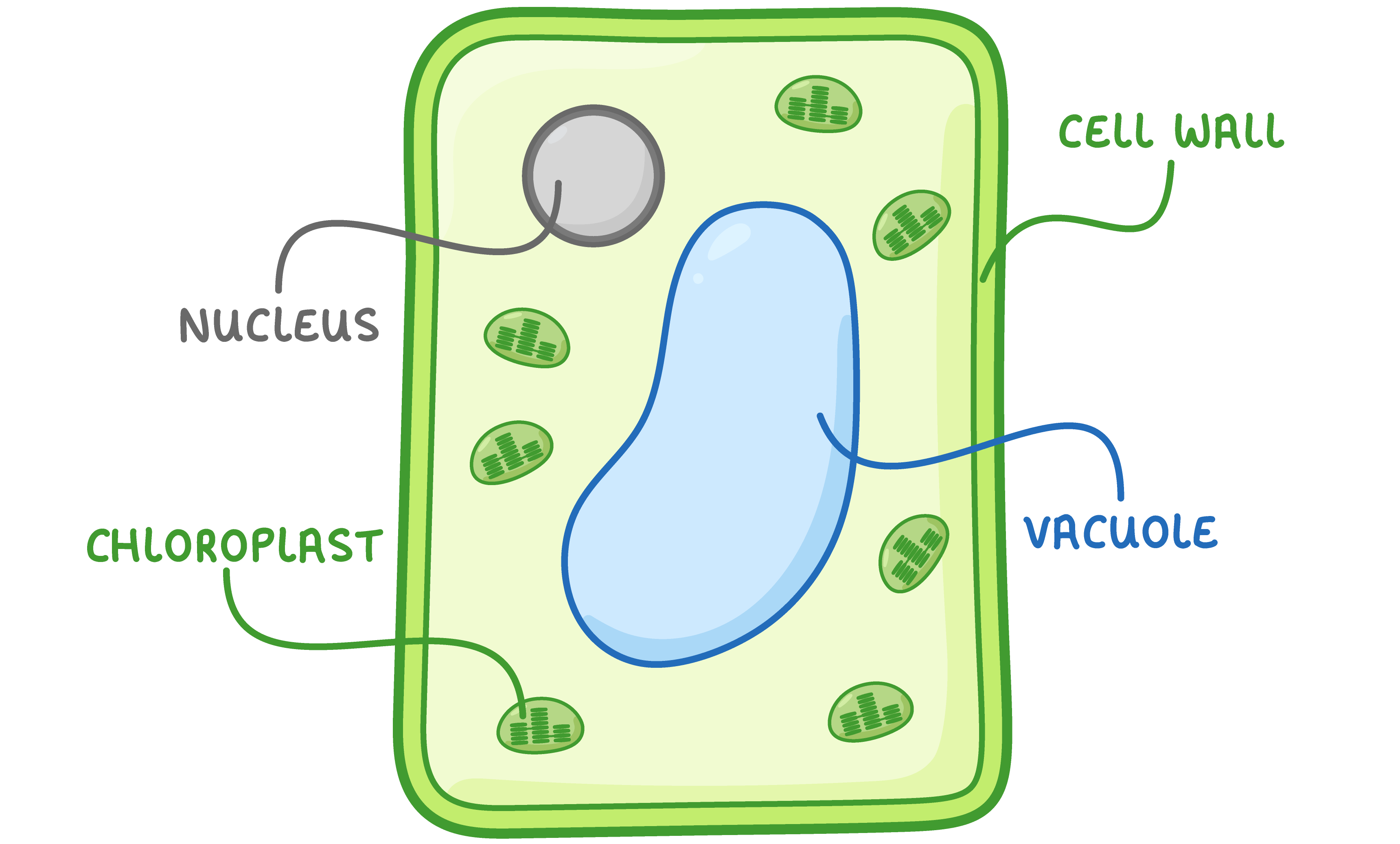
Palisade cells

Palisade cells carry out photosynthesis in the leaves of a plant. 


Specialised features: 

  • Lots of chloroplasts - These absorb the light needed for photosynthesis. 
  • Thin cell walls - This allows carbon dioxide to quickly diffuse into the cell. 
  • Tall and thin shape - This allows many palisade cells to closely pack together to form a continuous layer near the surface of the leaf. 
Diagram of a root hair cell showing mitochondria, cell wall, and root hair.

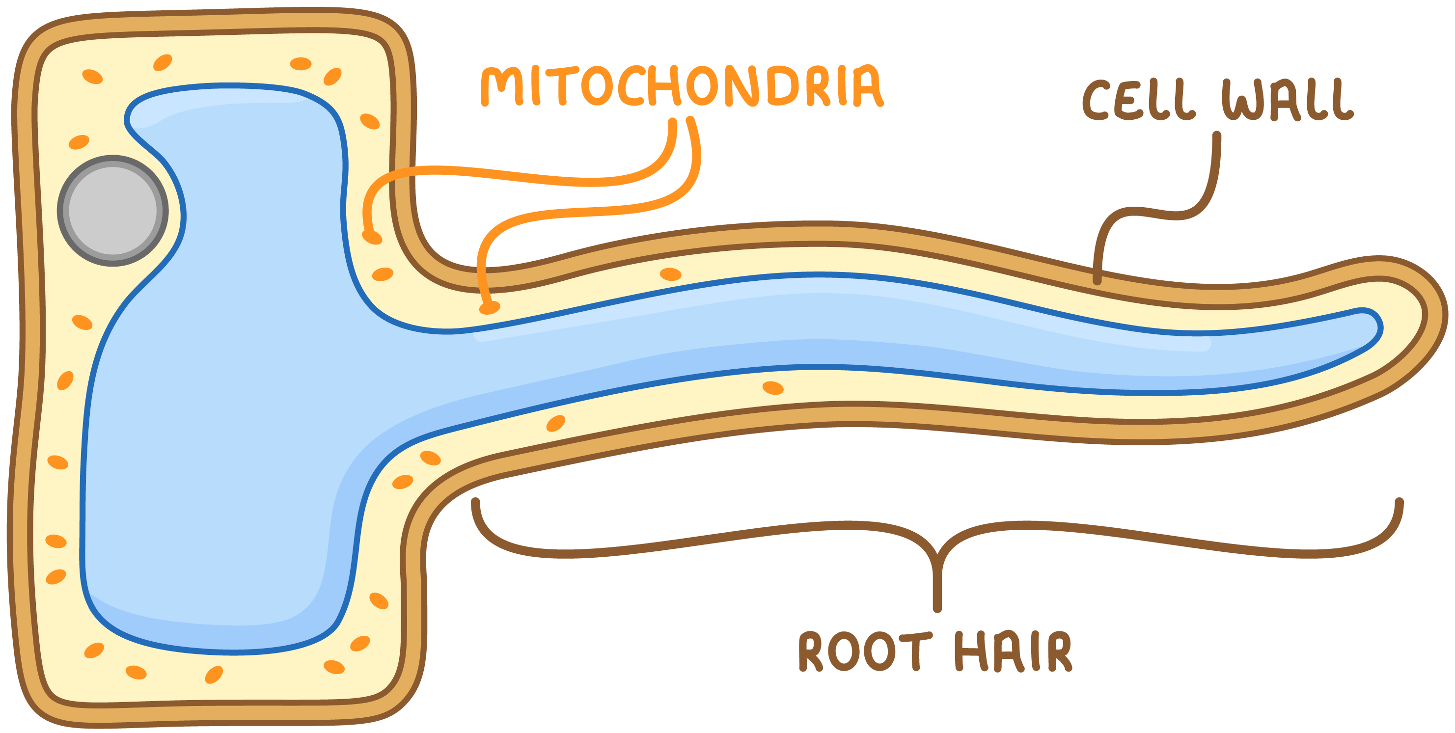
Root hair cells

Root hair cells absorb water and mineral ions from the soil. 


Specialised features: 

  • Root hair structures - These increase the surface area for absorption. 
  • Thin, permeable cell wall - This allows entry of water and ions. 
  • Lots of mitochondria - These provide energy for active transport. 
Illustration showing open and closed stoma with labels for thicker inner wall and thinner outer wall.

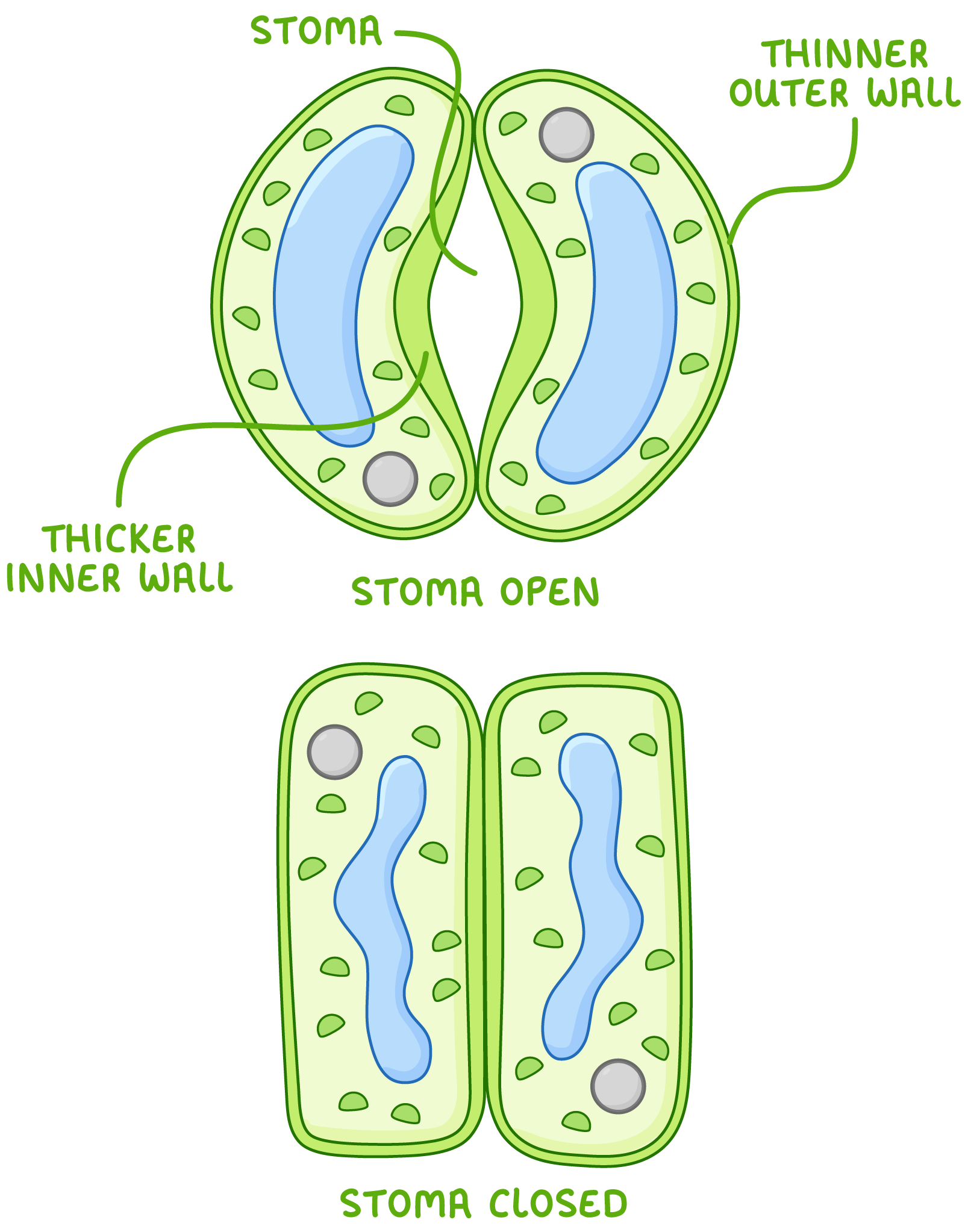
Guard cells 

Guard cells control the opening and closing of stomata. They are used to allow carbon dioxide to enter the leaves and to prevent water loss.


Specialised features: 

  • Come in pairs - This allows a gap (stoma, plural: stomata) to form between them. 
  • Change shape when light is present - Guard cells absorb water to become turgid, opening the stoma to allow entry of carbon dioxide. 
  • Change shape when they lose water - Guard cells shrink and close the stoma to prevent water loss. 
  • Thin outer walls and thick inner walls - This allows the cells to bend when they are turgid to open the stoma.