Fluid Mosaic Membranes

This lesson covers:

  1. The role of cell membranes in living organisms 
  2. The fluid-mosaic model of cell membranes
  3. The structure and function of cell membrane components 

Roles of cell membranes

There are two main types of cell membrane: 

  1. Cell-surface membranes - These surround cells to act as a barrier between the cell and its environment, controlling which substances enter and leave the cell. 
  2. Membranes around organelles - These surround organelles (e.g. mitochondria) to act as a barrier between the organelle and the cytoplasm, dividing the cell into different compartments (this is sometimes called compartmentalisation). 


Both types of membrane are partially permeable, meaning they let some molecules pass through, but not others. 

Fluid-mosaic model

In 1972, the fluid-mosaic model was proposed to describe the structure of cell membranes:

  • Fluid - Phospholipids form a bilayer in which the phospholipid molecules are constantly moving.
  • Mosaic - There are proteins of different sizes and shapes embedded in the phospholipid bilayer (so it looks a bit like a mosaic).

Structure of the membrane

Diagram showing the structure of a cell membrane with labelled components including phospholipid bilayer, cholesterol, intrinsic protein, extrinsic proteins, glycoprotein, and glycolipid.

Key components of cell membranes:

  • Phospholipid bilayer - This is made up of phospholipids with hydrophilic heads and hydrophobic tails.
  • Cholesterol - This adds stability.
  • Proteins - These may be intrinsic and extrinsic proteins.
  • Glycoproteins - These are proteins attached to a carbohydrate.
  • Glycolipids - These are lipids attached to a carbohydrate.

Phospholipid bilayer

Each phospholipid molecule is made up of a hydrophilic 'head' and a hydrophobic 'tail'.


This causes phospholipids to arrange themselves into a bilayer so that the hydrophilic heads are facing out (towards water) and the hydrophobic tails are facing in (away from water). 

Diagram showing the phospholipid bilayer with hydrophilic heads facing outwards and hydrophobic tails facing inwards.

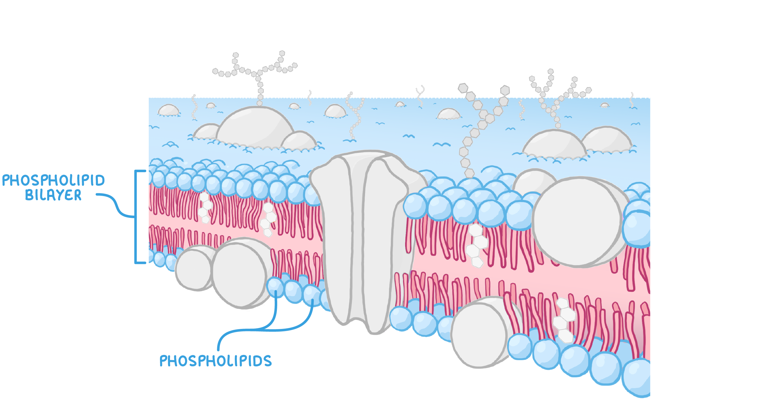
This arrangement creates a hydrophobic centre in the bilayer so that water-soluble substances cannot pass through. 


However, fat-soluble substances can dissolve in the bilayer and pass directly through the cell membrane. 

Cholesterol

Cholesterol molecules provide stability to cell membranes.

Illustration showing cholesterol molecules in a cell membrane, highlighting their hydrophilic and hydrophobic regions.

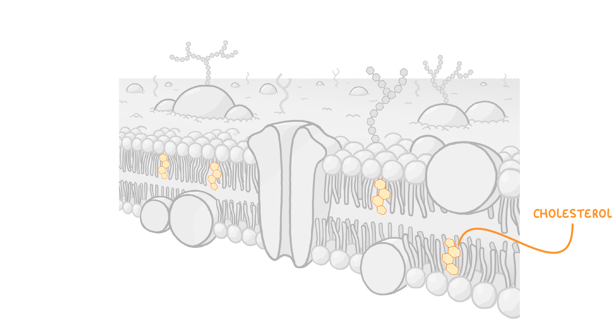
Cholesterol molecules consist of a hydrophilic and hydrophobic region. The hydrophobic regions bind to phospholipid fatty acid tails, causing them to pack more closely together.


This reduces the fluidity of the cell membrane. 

Proteins

Proteins in cell membranes can be grouped into two categories: intrinsic and extrinsic proteins. 
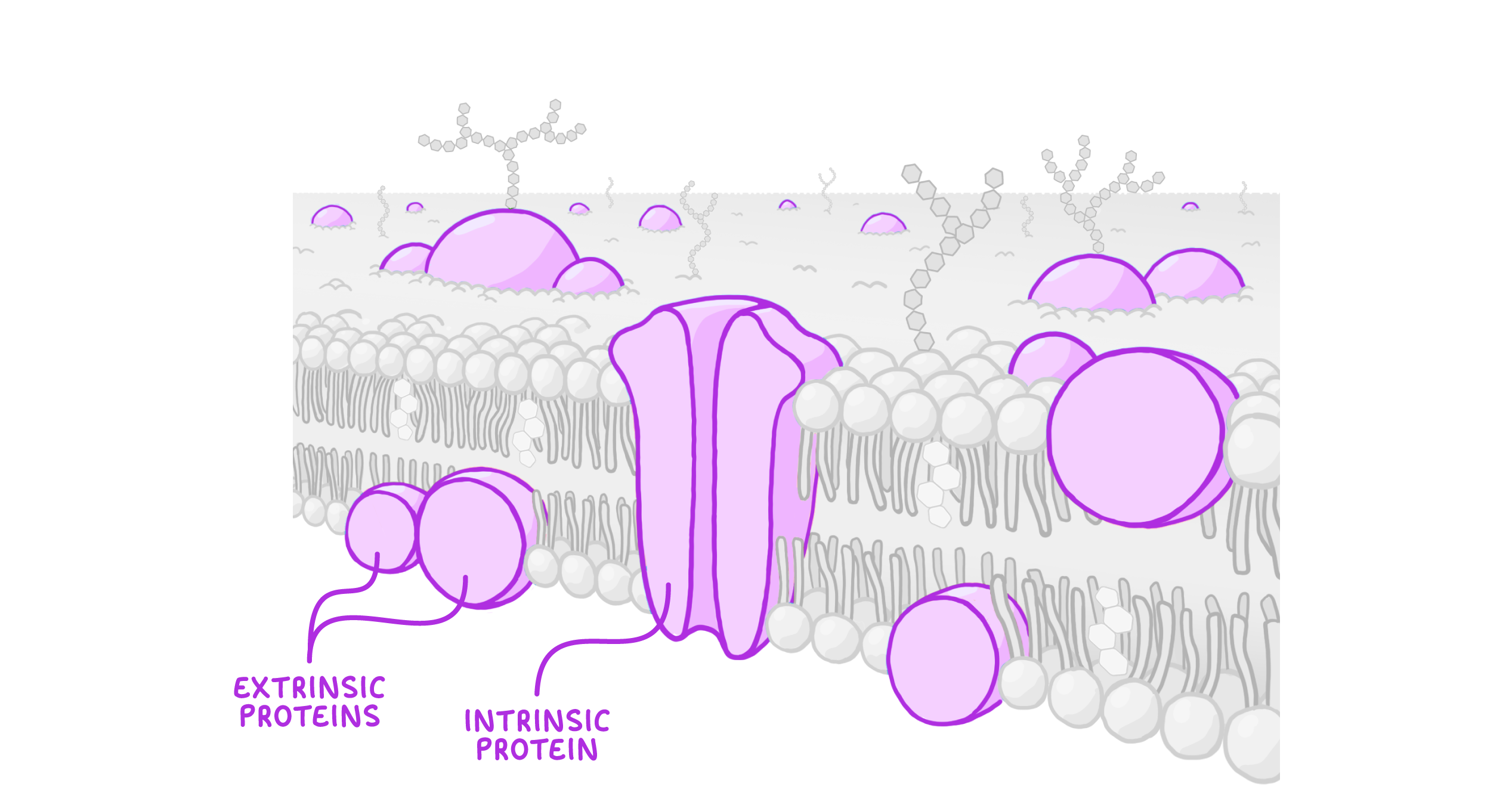
Illustration showing intrinsic and extrinsic proteins in a cell membrane.

Intrinsic proteins:

  • Intrinsic (or integral) proteins are embedded through both sides of the phospholipid bilayer.
  • These proteins include channel and carrier proteins which transport large molecules and ions across the membrane.


Extrinsic proteins:

  • Extrinsic (or peripheral) proteins are present on only one side of the phospholipid bilayer.
  • These proteins provide support to the membrane or may be involved in cell signalling. 

Glycoproteins and glycolipids

Glycoproteins consist of intrinsic proteins attached to carbohydrates, whereas glycolipids consist of lipids attached to carbohydrates. 

Illustration of glycoproteins and glycolipids in a cell membrane, showing cell adhesion, cell recognition, and cell signalling.

Glycoproteins and glycolipids are involved in the following: 

  1. Cell adhesion - This is the attachment of cells to one another. 
  2. Cell recognition - This allows cells to recognise one another.
  3. Cell signalling - This is communication between cells.