Meiosis
This lesson covers:
- What 'meiosis' is
- The stages of meiosis
- The differences between meiosis and mitosis
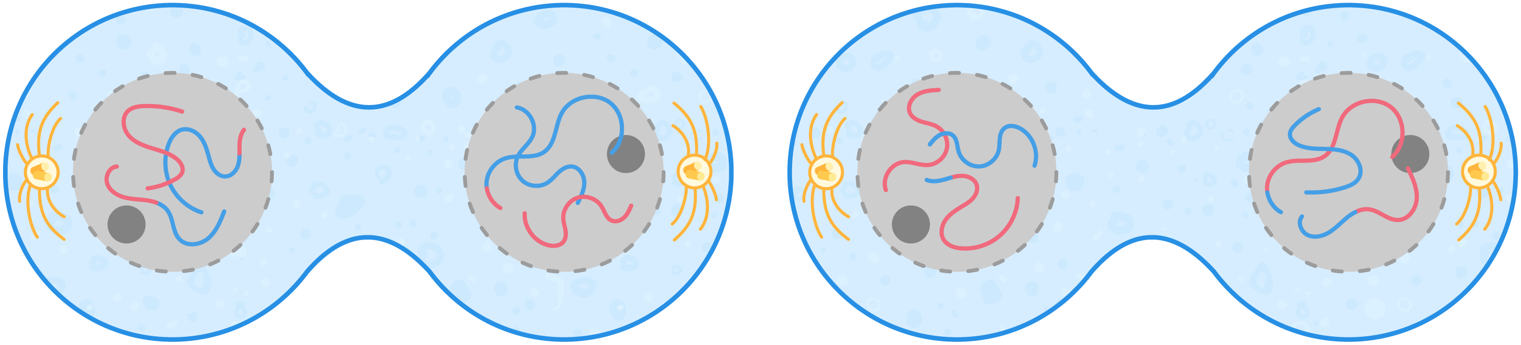
Overview of meiosis Meiosis is a type of cell division in which a parent cell divides to form four haploid cells, each genetically distinct from one another. Before meiosis starts, DNA is replicated during interphase so that each chromosome contains two chromatids. 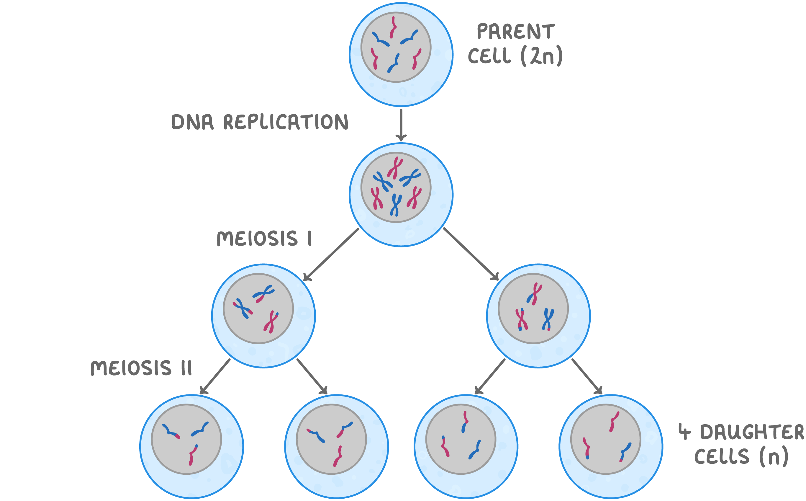 |
The cell then undergoes 2 divisions:
Meiosis involves a reduction division in which the chromosome number is halved to form haploid cells. These haploid cells form the gametes (egg and sperm cells) in animals and plants. |
Stages of meiosis The two divisions of meiosis contain the same stages as mitosis:
Remember that cells have already replicated their DNA during interphase and each chromosome is made up of two identical sister chromatids. |
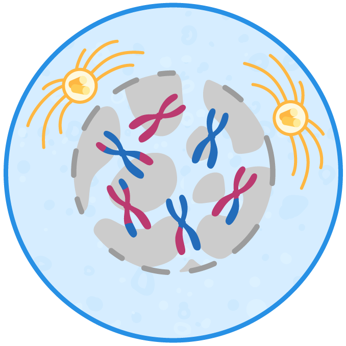 Prophase I The chromosomes condense and homologous chromosomes pair up. Centrioles migrate to opposite poles of the cell where each centriole starts forming spindle fibres. The nucleolus disappears and the nuclear envelope starts to break down, leaving the chromosomes free in the cytoplasm. |
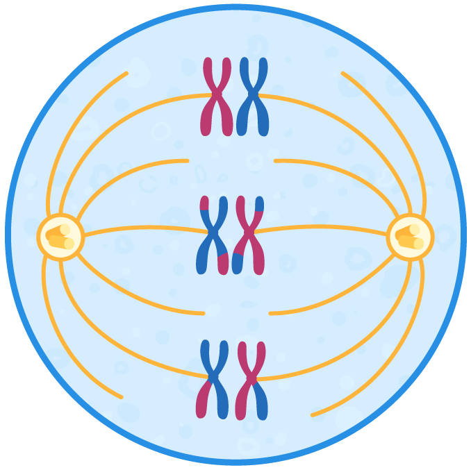 Metaphase I Chromosomes line up along the equator of the cell in their homologous pairs (so in humans, 23 pairs line up). Each chromosome attaches to the spindle by their centromere. |
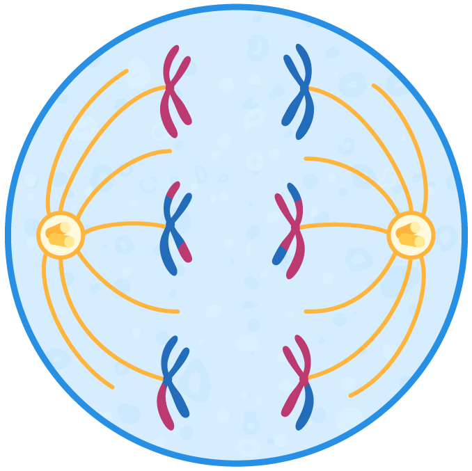 Anaphase I Homologous chromosome pairs are separated and pulled to opposite poles of the cell (chromatids stay joined together). |
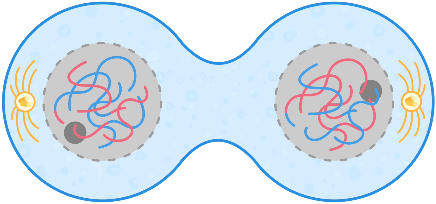 Telophase I The chromosomes reach the opposite poles of the cell where they uncoil. A nuclear envelope forms around each set of chromosomes and the nucleolus starts to reform. The cytoplasm divides to form two cells (cytokinesis). |
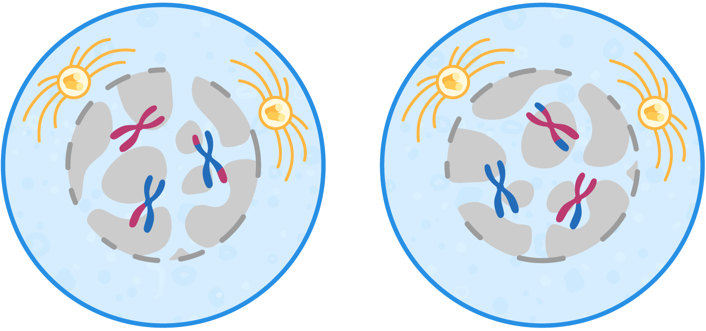 Prophase II The chromosomes condense and are now visible under a microscope. Centrioles migrate to opposite poles of the cell where each centriole starts forming spindle fibres. The nucleolus disappears and the nuclear envelope starts to break down. |
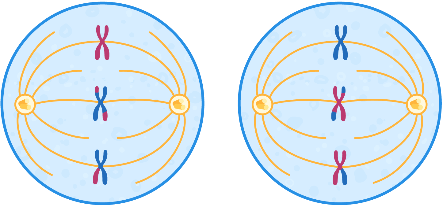 Metaphase II The chromosomes line up at the equator of the cell (so in humans, 23 chromosomes line up). Each chromosome attaches to the spindle by their centromere. |
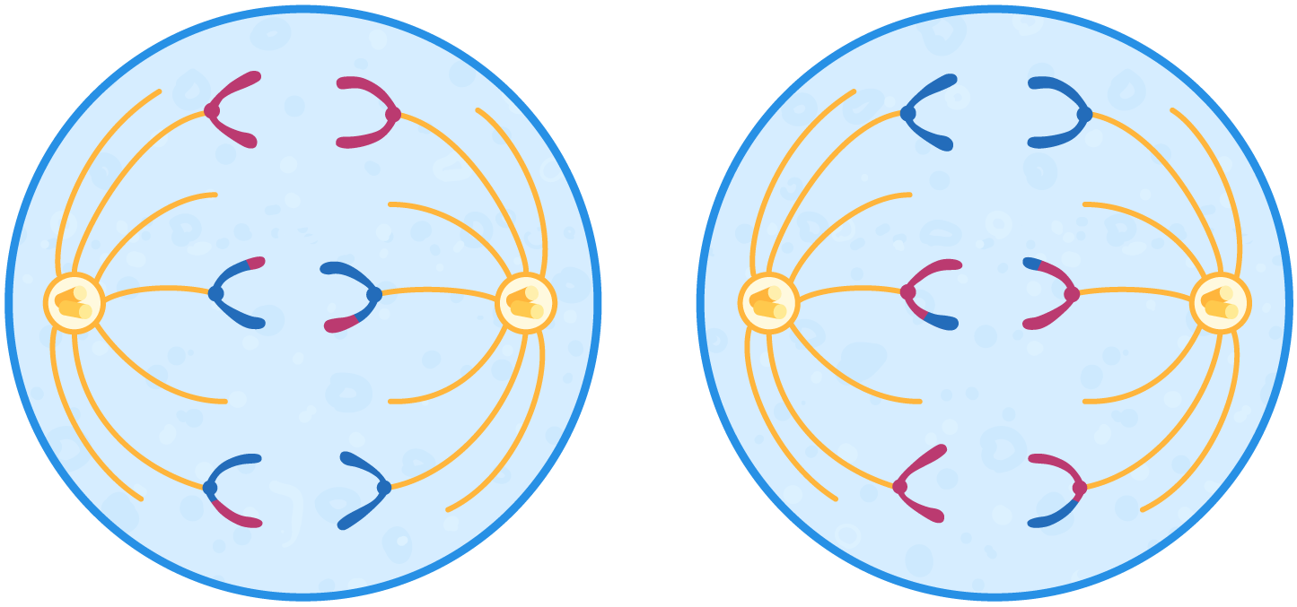 Anaphase II The centromeres divide and separate each pair of chromatids. The spindle fibres contract and shorten to pull the chromatids to opposite poles of the cell. |
 Telophase II The chromatids reach the opposite poles of the cell where they uncoil to become long and thin again. A nuclear envelope forms around each set of chromosomes to form two nuclei and the nucleolus starts to reform. The cytoplasm divides (cytokinesis) and 4 cells are produced. |
Comparing mitosis and meiosis 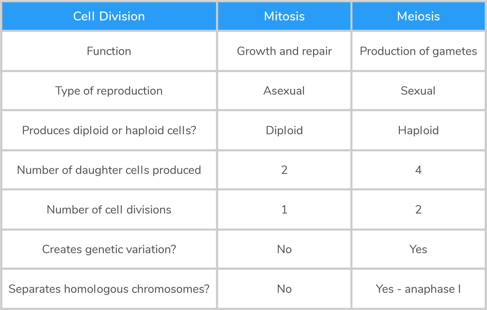 |