Electron Microscopes
This lesson covers:
- How a transmission electron microscope works
- How a scanning electron microscope works
- The differences between light and electron microscopes
Electron microscopes Electron microscopes use electrons rather than light to form an image. Since electrons have a shorter wavelength than light, electron microscopes have a higher resolution than light microscopes. However, electron microscopes are more expensive than light microscopes and only produce images in black and white (computers can add colour to these images). The two types of electron microscope you need to know are:
These microscopes require complex preparation of specimens, meaning they are more likely to create artefacts. Artefacts are visible details that aren't part of the specimen being observed, such as air bubbles or fingerprints. |
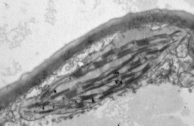
Transmission electron microscopes Transmission electron microscopes (TEMs) use electromagnets to transmit a beam of electrons through a specimen. The denser parts absorb more electrons, so appear darker in the image formed. TEMs produce high resolution images, meaning they can be used to observe the internal structures of organelles (e.g. chloroplasts). The diagram below shows a chloroplast as viewed under a TEM. |
 |
However, the specimen must be viewed in a vacuum, meaning only non-living or dead organisms can be observed. Also, the specimen must be thin to allow electrons to pass through. TEMs have a higher resolution and magnification compared to light microscopes:
|
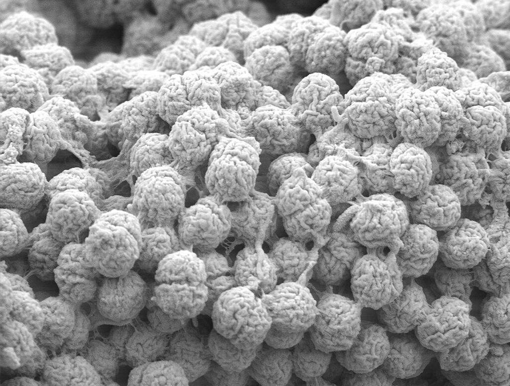
Scanning electron microscopes Scanning electron microscopes (SEMs) scan a beam of electrons across the surface of a specimen. Reflected electrons are then used to form an image. SEMs produce 3D images of the surface of the specimen. The diagram below shows algae as viewed under a SEM. |
 |
Like TEMs, SEMs can only view non-living or dead specimens. However, SEMs can be used on thicker specimens. SEMs have a lower resolution and similar magnification compared to TEMs:
|
Comparing light and electron microscopes
| Microscope | Light | TEM | SEM |
|---|---|---|---|
| Magnification | ×1,500 | ×1,500,000 | ×1,500,000 |
| Resolution | 0.2 µm | 0.5 nm | 5 nm |
| Cost | Inexpensive | Expensive | Expensive |
| Sample preparation | Simple | Complex | Complex |
| Image produced | 2D, colour | 2D, black and white | 3D, black and white |
| Specimens used | Living or dead | Dead | Dead |