Gas Exchange in Insects

This lesson covers:

  1. Why insects need gas exchange systems
  2. Structural and functional compromises in insect gas exchange
  3. The structures involved in insect gas exchange
  4. How gas exchange occurs in insects
  5. Additional ventilation mechanisms in insects

Why insects need gas exchange

Insects have high oxygen demands but their tough chitinous external skeleton (exoskeleton) prevents direct gas exchange.


Insects need efficient systems for exchanging gases for two main reasons:

  1. To deliver oxygen to cells - This allows aerobic respiration to occur to release energy for cellular processes.
  2. To remove carbon dioxide from cells - The build up of carbon dioxide produced as a waste product of respiration reduces pH, which can denature enzymes.

Insect gas exchange systems have adapted to balance two conflicting needs:

  1. Maximising gas exchange efficiency
  2. Minimising water loss


The exoskeleton is covered with a waterproof cuticle to help prevent water loss, but the insect gas exchange system itself has additional methods to prevent excess water loss while still being effective at gas exchange.

Structures of the insect gas exchange system

Insects have an open respiratory system comprised of tubular structures that transport air.

Diagram showing the structures of the insect gas exchange system including spiracle, tracheae, tracheoles, and tracheal fluid.

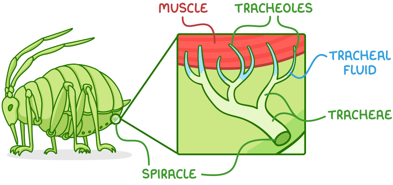
The main structures involved are:

  1. Tracheae - These are air-filled tubes branching throughout the body.
  2. Tracheoles - These are fine branches of tracheae that deliver gases to cells.
  3. Spiracles - These are external openings of the tracheal system on the exoskeleton along the abdomen and thorax.

Adaptations of the structures in the insect gas exchange system

There are some key features that make these structures well adapted for their functions.


Tracheae:

  • Reinforced with spirals of chitin - This prevents collapsing.
  • Multiple tracheae - This increases surface area.

Tracheoles:

  • Penetrate directly into tissues - This reduces the gas diffusion distance.
  • Thin walls - These reduce the gas diffusion distance.
  • Highly branched - This maximises the surface area.
  • Not reinforced with chitin - This allows gas exchange to occur.
  • Fluid at the ends of the tracheoles (tracheal fluid) - This allows oxygen to dissolve to aid diffusion and reduces water loss.

Spiracles:

  • Open and close - This allows them to control gas exchange with the atmosphere and minimise water loss.

How gas exchange occurs

There are several key stages to gas exchange in insects.
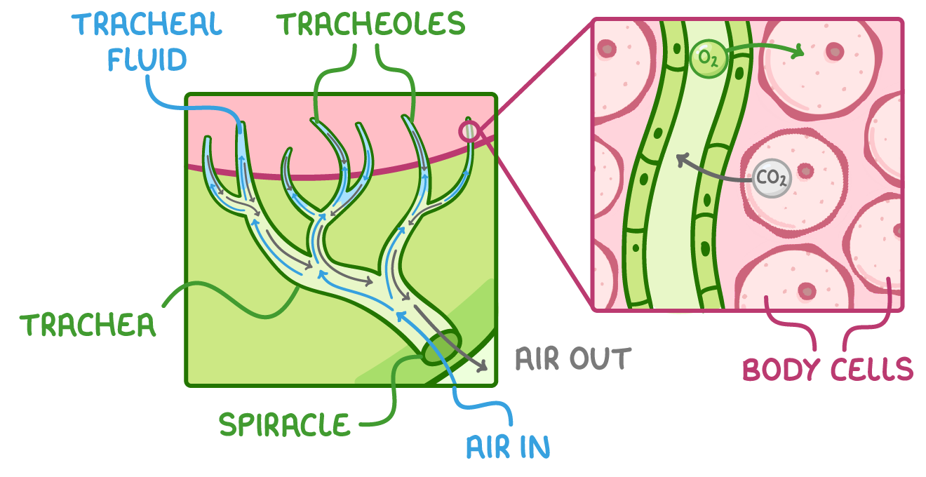
Diagram showing gas exchange in insects with trachea, tracheoles, spiracle, tracheal fluid, air in, air out, and body cells.

These stages are:

  1. Air enters the tracheal system through open spiracles.
  2. Air moves into larger tracheae and diffuses into smaller tracheoles.
  3. Tracheoles branch throughout the body, transporting air directly to cells.
  4. Oxygen dissolves in water in tracheal fluid and diffuses down its concentration gradient from tracheoles into body cells.
  5. Carbon dioxide diffuses down its concentration gradient out of body cells into the tracheoles.
  6. Air is then carried back to the spiracles via the tracheae and released from the body.

The concentration gradients between the tissues and air in the tracheal system are maintained by:

  1. Cells using up oxygen for respiration - This keeps oxygen concentration low in cells.
  2. Cells producing carbon dioxide in respiration - This keeps carbon dioxide concentration high in cells.
  3. Continuous ventilation - Fresh air is supplied to the tracheal system via spiracles.

Other ventilation mechanisms

Some insects, particularly active ones, may use additional ventilation mechanisms to drive air through the tracheal system.


Some of these mechanisms include:

  1. Mechanical active ventilation - This is when muscles around the tracheae contract and relax, changing the volume and pressure in the abdomen and squeezing the tracheae to pump air in and out of the spiracles.
  2. Tracheal fluid - This moves out into tissues during exercise to increase the diffusion rate and surface area for gas exchange.
  3. Enlarged collapsible tracheae, or accessory sacs and air reservoirs - These inflate or deflate to ventilate the tracheal system and can increase the volume of air moved through the system.
  4. Wing muscles connected to sacs - These pump air to ventilate the tracheal system.
  5. Vibration of thoracic muscles - This pumps air to ventilate the tracheal system.