Gel Electrophoresis
This lesson covers:
- What gel electrophoresis is
- Running electrophoresis on DNA samples
- Visualising and analysing results from gel electrophoresis
What is gel electrophoresis?
Gel electrophoresis is a technique used to separate molecules such as DNA, RNA, or proteins based on size by using an electric current applied to an agarose gel matrix.
How to set up gel electrophoresis:
- Insert a gel tray with solidified agarose gel into a gel tank.
- Ensure the wells are close to the negative electrode to position the gel correctly.
- Pour a buffer solution over the gel until it is submerged to maintain a constant, suitable pH throughout the experiment.
Running electrophoresis on DNA samples
To run gel electrophoresis, first the samples need to be loaded into wells.
To load samples:
- Mix the DNA samples with loading dye to make them visible.
- Carefully deposit equal volumes of each sample into the wells using a micropipette.
- Touch the micropipette tip to the buffer, not the bottom of the gel, to prevent damaging the gel.
- Keep a record of which sample is in each well for later analysis.
DNA molecules carry a negative charge due to their phosphate groups.
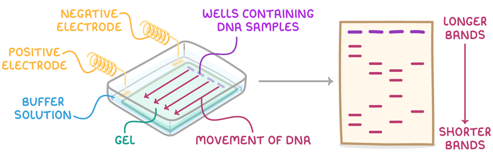
During electrophoresis:
- A voltage (around 100 V) is applied across the gel.
- Fragments of DNA move towards the positive electrode (anode).
- The smaller fragments travel faster and thus separate by size.
- Continue the process for about 30 minutes or until the dye approaches the end of the gel.
The size-based separation occurs because the gel's mesh matrix slows down larger fragments more than the smaller ones. The pore size of the gel affects the rate at which fragments move.
Visualising and analysing the results from gel electrophoresis
There are a few steps needed to visualise the results form gel electrophoresis.
To visualise the results:
- Switch off the voltage and remove the gel from the tank.
- Apply a stain to the DNA to reveal the bands of fragments.
- Assess the migration distances of the bands to approximate the sizes of the fragments.