Cell Specialisation & Organisation
This lesson covers:
- The organisation of cells into tissues, organs, and organ systems
- Examples of the structures and functions of different tissues
Key terms
|
Levels of organisation The organisation of cells in living organisms can be summarised as follows: 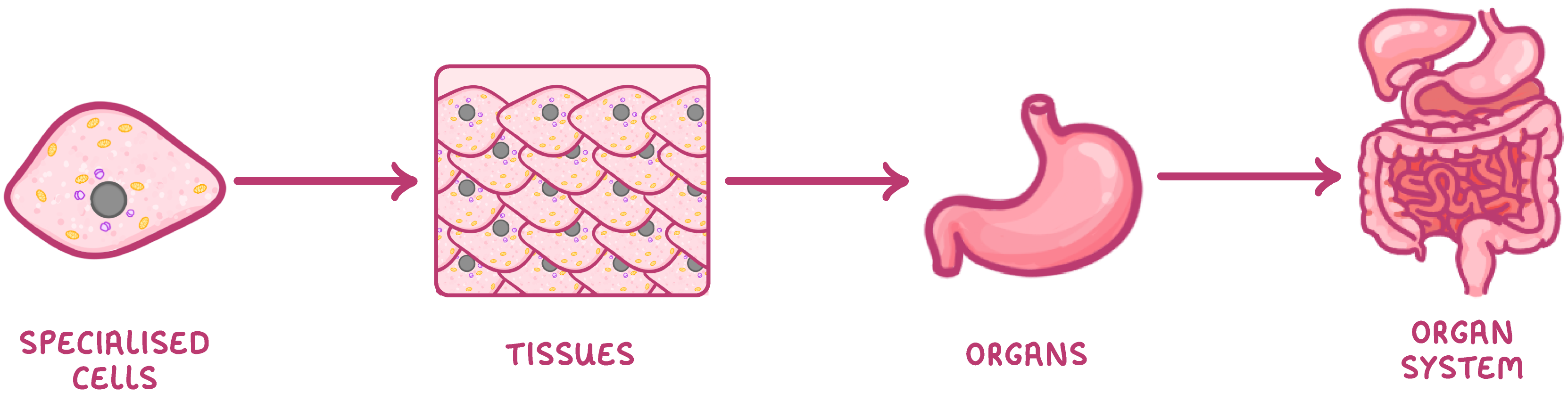 |
Examples of these levels include:
|
Examples of animal tissues You need to know about the following types of animal tissues. |
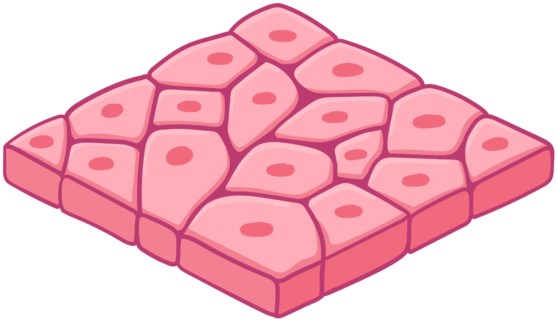 Squamous epithelium Squamous epithelium tissue provides a thin lining for many organs such as the lungs. This tissue is made up of a single layer of squamous epithelial cells. Due to being only one cell thick, gases can quickly diffuse through the tissue. |
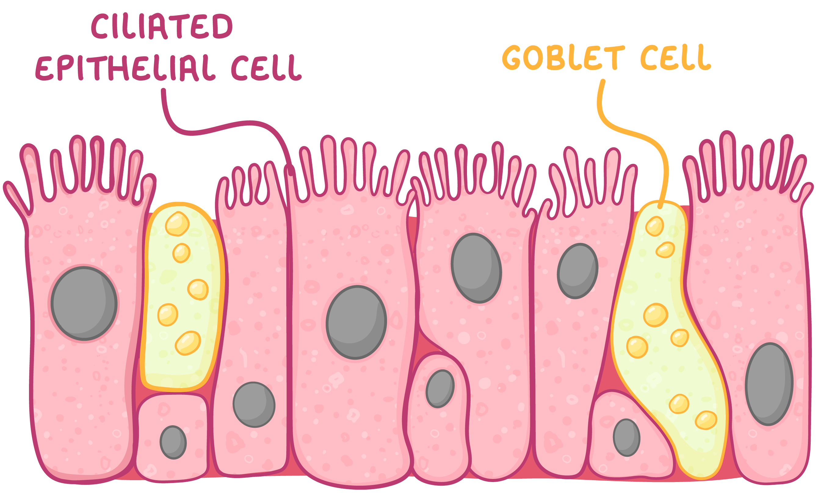 Ciliated epithelium Ciliated epithelium tissue lines organs such as the trachea where it can sweep mucus away from the lungs. This tissue is made up of ciliated epithelial cells and goblet cells. The goblet cells release mucus to trap pathogens, whilst the ciliated epithelial cells use cilia to sweep the mucus away. |
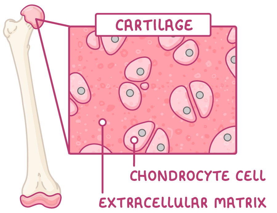 Cartilage Cartilage is a type of connective tissue that acts as a cushion between bones and also provides support to organs such as the ears and nose. This tissue is made up of chondrocyte cells fixed within an extracellular matrix. |
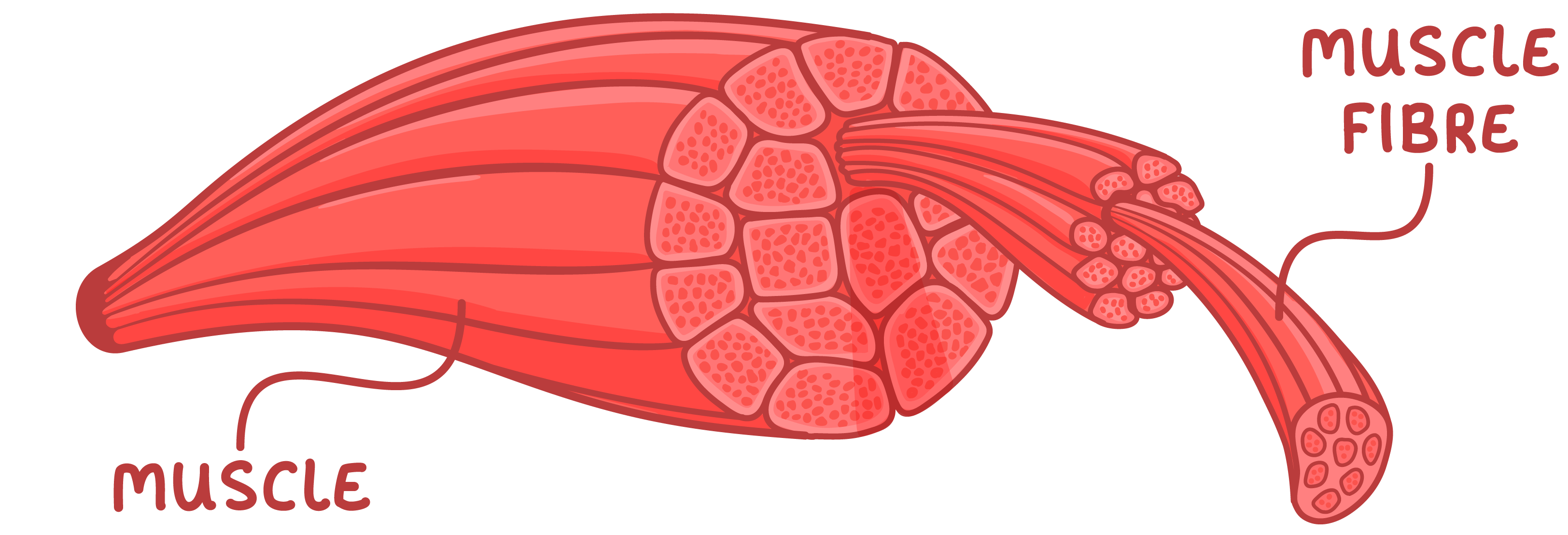 Muscle Muscle tissue is made up of muscle fibres (bundles of elongated cells). These fibres contract (shorten) and relax to move different parts of the body. There are three types of muscle tissue:
|
Examples of plant tissues You need to know about the following types of plant tissues. |
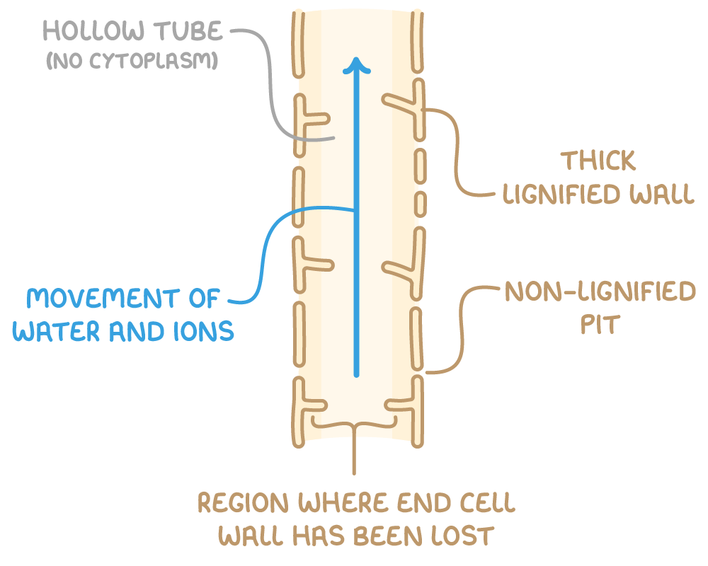 Xylem Xylem tissue is responsible for the transport of water and minerals within plants. It is made up of dead xylem vessel cells which have no end walls and no organelles. This forms a continuous column through which water can flow. The walls of these cells are strengthened by a waterproof material known as lignin. |
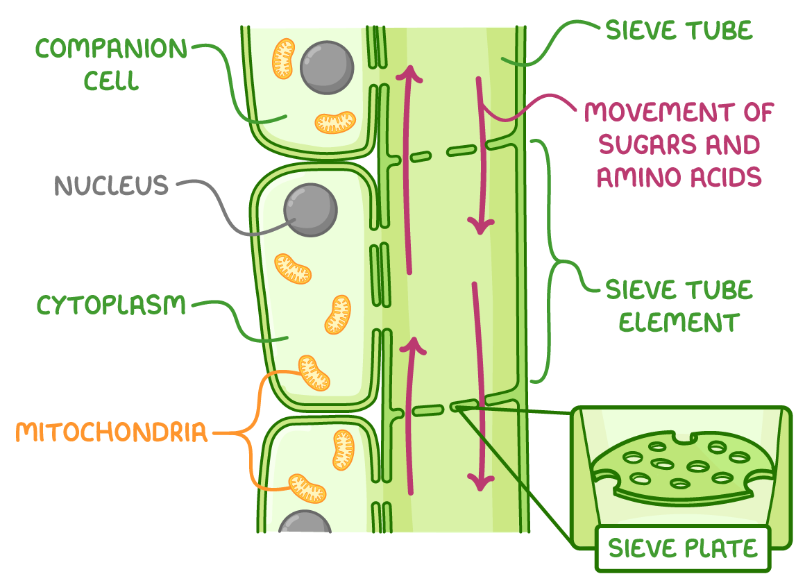 Phloem Phloem tissue is responsible for the transport of sugars and amino acids within plants. It is made up of columns of sieve tube elements and companion cells. The sieve tube element cells are separated by sieve plates with holes so that sugars can pass through. Sieve tube elements contain very few organelles, allowing sugars to flow easily. Companion cells contain many mitochondria to release energy. |