Cell Fractionation
This lesson covers:
- The purpose and process of cell fractionation
- The steps of cell fractionation
What is cell fractionation?
Cell fractionation is a laboratory method used to isolate different components of the cell, specifically the organelles, so they can be studied in detail, such as under an electron microscope.
This technique involves breaking the cell open and then separating the organelles based on their size and density.
The steps of cell fractionation
There are four main steps in cell fractionation:
- Sample preparation
- Homogenisation
- Filtration
- Ultracentrifugation
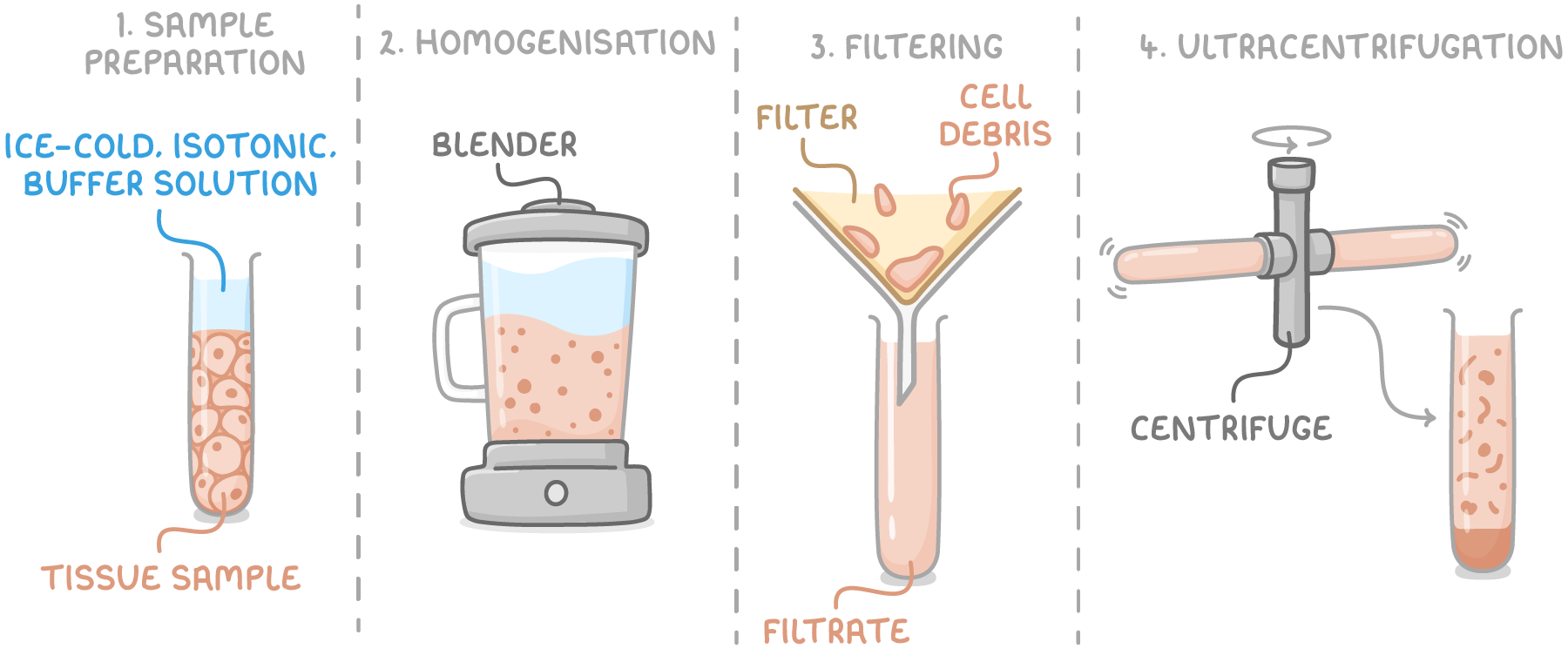
Step 1 - Sample preparation
The initial step in the cell fractionation process is to prepare the tissue sample. This involves placing the sample in an ice-cold, isotonic, buffered solution to protect the organelles during the fractionation process.
The solution used in sample preparation has the following characteristics:
- Ice-cold - This slows down enzyme activity that might otherwise break down organelles.
- Isotonic - It ensures that the water potential inside and outside the organelles is the same, preventing damage through water movement.
- Buffered - Keeping the pH constant is crucial to prevent denaturation of proteins and enzymes.
Step 2 - Homogenisation
The homogenisation step involves physically breaking open the cells. This disrupts the plasma membrane, which allows the organelles to be released into the solution.
This can be achieved using various methods, such as using a blender to grind the cells.
Step 3 - Filtration
Following homogenisation, the mixture is then filtered to remove larger pieces of cell debris and any remaining tissue fragments.
The filtration is typically done through a gauze, which allows smaller organelles to pass through while retaining larger debris.
Step 4 - Ultracentrifugation
Ultracentrifugation is the process used to separate organelles based on their density. By spinning the filtered solution at various speeds, organelles are separated into layers according to their mass.
Key definitions:
- Pellet - The sediment at the bottom of the tube, containing the heavier organelles.
- Supernatant - The liquid remaining above the pellet, which contains the lighter organelles.
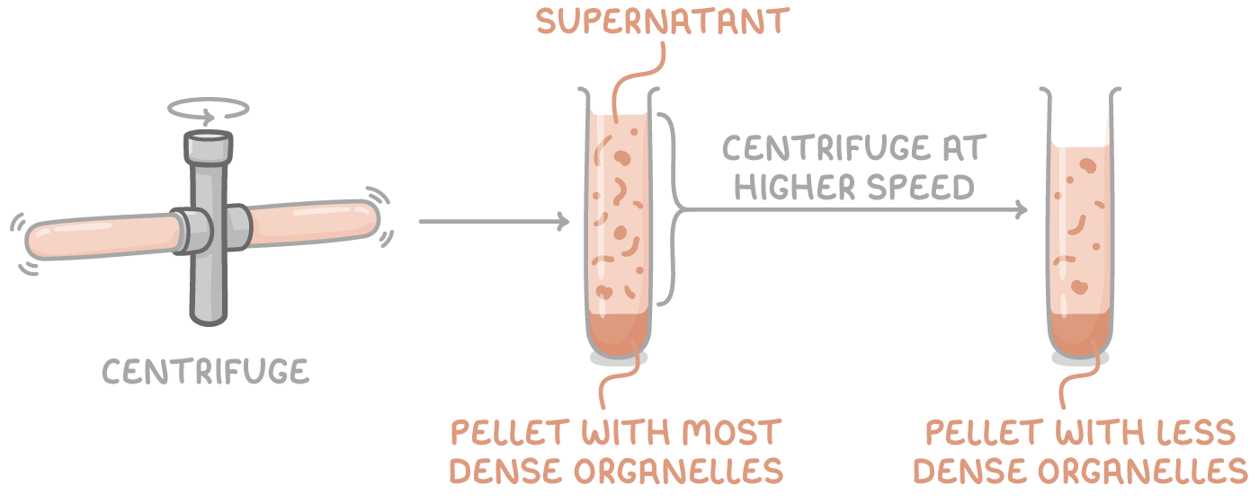
The ultracentrifugation process:
- The cell fragments are placed in a centrifuge tube and spun at a low speed.
- This results in the heaviest organelles, such as nuclei, forming a pellet at the bottom of the tube.
- The lighter organelles remain suspended in the supernatant.
- The supernatant is then transferred to a new tube and centrifuged at a higher speed.
- This leads to the next heaviest set of organelles, typically mitochondria, settling into a pellet.
- Steps 4 to 5 are repeated, increasing the speed each time to separate the remaining organelles until all organelles have been separated into distinct layers.
The order of organelles from heaviest to lightest:
- Nuclei (heaviest)
- Chloroplasts
- Mitochondria
- Lysosomes
- Endoplasmic reticulum
- Ribosomes (lightest)