Ventilation
This lesson covers:
- What ventilation is
- The muscles involved in breathing
- The process of inspiration
- The process of expiration
What is ventilation? Ventilation, or breathing, is the constant movement of air into and out of the lungs. It consists of inspiration (breathing in) and expiration (breathing out). It allows air to enter and leave the lungs, providing the body with oxygen and removing carbon dioxide. |
Muscles involved in ventilation The ribcage is made up of bones called ribs that enclose the thorax - the cavity where the lungs are located (thoracic cavity). In mammals, ventilation is controlled by specific muscles. When the muscles attached to the ribcage contract and relax, they move the ribs to change the volume of the thoracic cavity. This affects the pressure in the lungs and controls ventilation. |
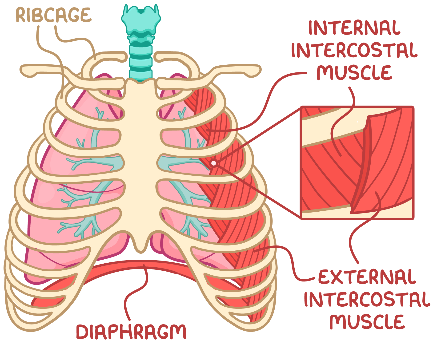 There are three sets of muscles that act on the ribcage:
|
The external and internal intercostal muscles have opposite effects on the ribcage. The external muscles expand the ribcage during inspiration, while the internal muscles shrink it during expiration. |
Inspiration Inspiration is an active process requiring energy for muscle contraction. |
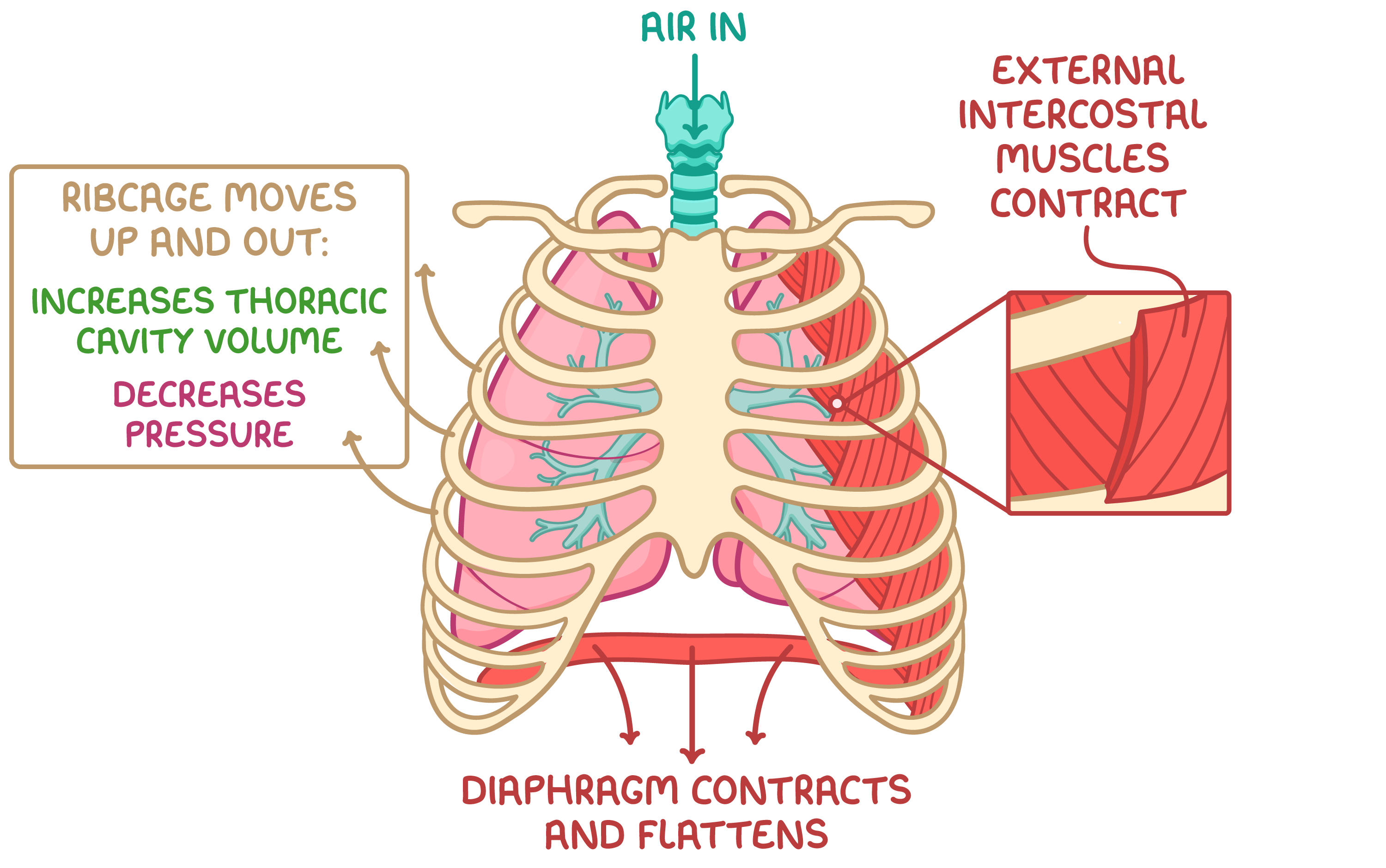 During inspiration:
|
Expiration Normal expiration at rest is a passive process so it does not require energy. However, expiration can be forced by contracting the internal intercostal muscles to actively pull the ribcage down and in, forcing more air out. |
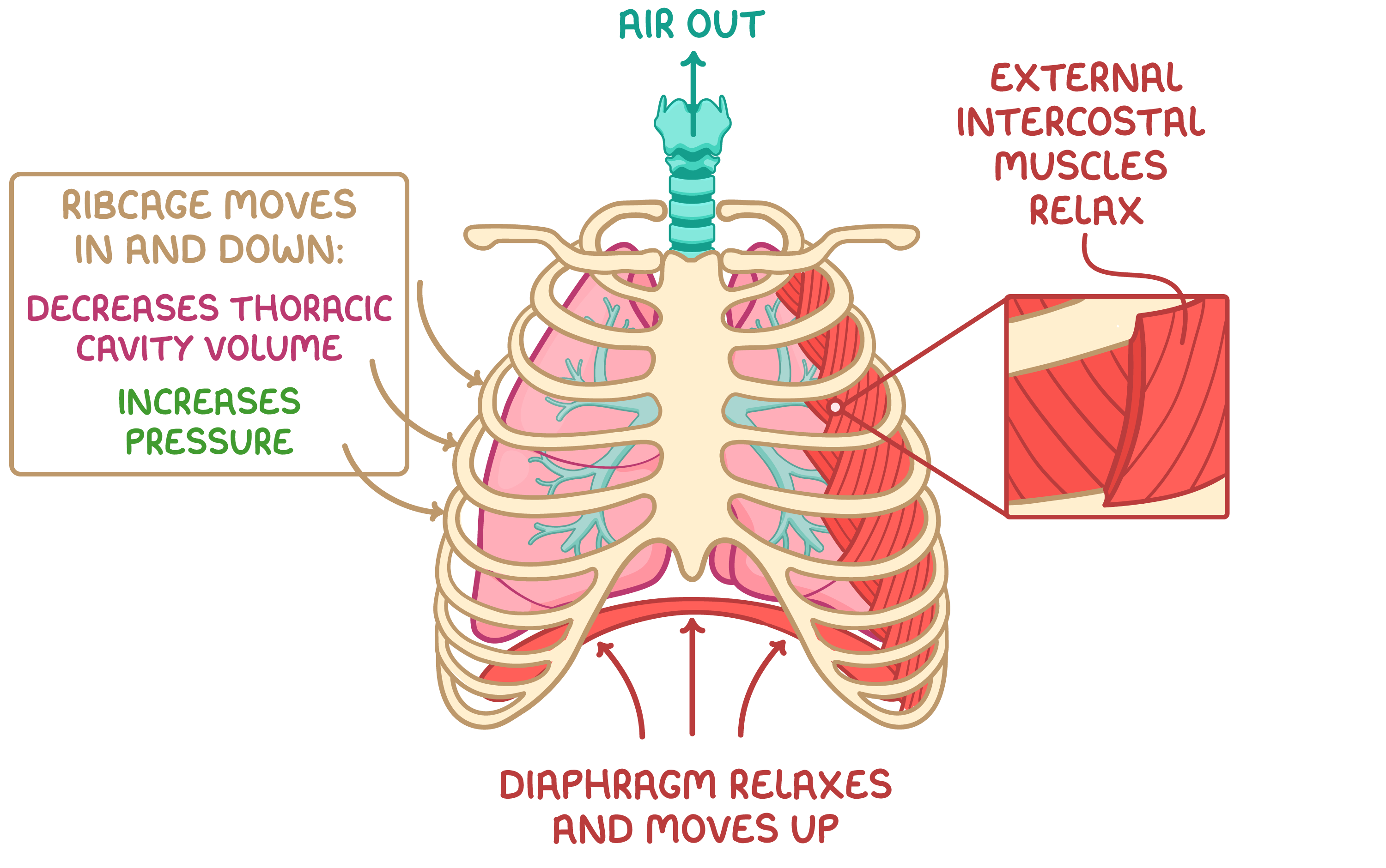 During expiration:
|
Elastic fibres in the alveoli also shrink and recoil back to their original shape when the thoracic cavity volume decreases. This increases the pulmonary pressure and helps to push air out of the lungs. |