Photoreceptors
This lesson covers:
- How light enters the eye and reaches the photoreceptors
- How light is converted into an electrical signal by photoreceptors
- The key features of rods and cones
How light enters the eye
When light first enters the eye, it travels through several structures before our brains can process an image.
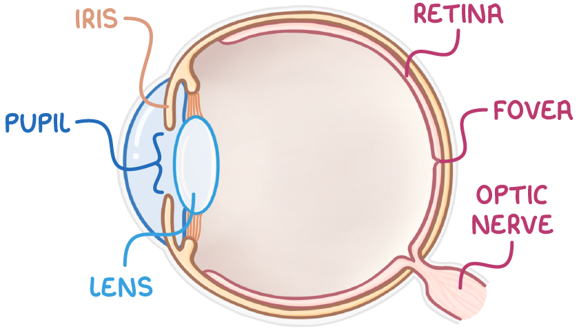
These key structures in the eye include:
- The pupil - The adjustable opening that lets light into the eye.
- The iris - The coloured part around the pupil that adjusts its size.
- The lens - The clear part that focuses light onto the back of the eye.
- The retina - The inner lining at the back of the eye, which contains photoreceptors.
- Photoreceptors - Cells that detect light.
- The fovea - An area in the retina full of cone cells for sharp central vision.
- The optic nerve - The nerve that sends signals from the retina to the brain.
How light is converted into an electrical impulse
Photoreceptors convert light into an electrical impulse.
This occurs in several stages:
- Light hits photopigments, which are light-sensitive proteins in photoreceptor cells.
- Light bleaches and causes a chemical change in the photopigments.
- This increases the permeability of photoreceptor cells to sodium ions.
- An influx of sodium ions generates a receptor potential.
- If this reaches threshold, a bipolar sensory neurone carries a signal to the optic nerve and then the brain.
The key features of rods and cones
There are two types of photoreceptor cell in the retina: rods and cones. They each contain different photopigments that together let our eyes work well in all sorts of light conditions.
| Feature | Rods | Cones |
|---|---|---|
| Pigment | Contain rhodopsin pigment, which makes rods sensitive to dim light | Contain iodopsin pigments, which makes cones sensitive to bright light of different wavelengths |
| Type of vision | Provide black-and-white vision | 3 different types of cone cell provide colour vision |
| Location in retina | Located in the peripheral retina, allowing for peripheral vision in low-light conditions | Densely packed in the fovea, because they require higher light intensities |
| Connection to neurones | Many rods converge on one bipolar neurone to sum weak signals in low light, increasing the chance of reaching threshold potential (retinal convergence) | Each cone has its own bipolar neurone, so more light is needed to trigger an impulse |
| Light sensitivity | Very light sensitive, detecting light at low intensities | Less sensitive than rods, only detecting light at high intensities |
| Visual acuity | Low visual acuity, as signals from multiple rods merge in one bipolar neurone, so two close light points appear as one | High visual acuity, as one-to-one connections between cone cells and bipolar neurones allow discrimination of fine details |
The number of rods and cones that connect to a single bipolar sensory neurone differs. This means their visual acuity also differs, which is a term for the sharpness of vision and ability to distinguish fine details.