Human Gas Exchange System

This lesson covers:

  1. An introduction to the gas exchange system
  2. The structure and function of the ciliated epithelium
  3. The structure and adaptations of the trachea, bronchi and bronchioles
  4. The structure and adaptations of the alveoli

Introduction to the gas exchange system

The human gas exchange system consists of the lungs and air passages. This system allows oxygen to enter the blood and carbon dioxide to leave the blood through gas exchange surfaces called alveoli. The lungs are located inside the chest in the thoracic cavity (thorax), protected by the ribcage.


The gas exchange system is located inside the body because:

  1. Air is not dense enough to support and protect these delicate structures.
  2. The body would otherwise lose water and dry out.

Pathway of air

Air travels through several structures as it passes through the respiratory system.

Diagram showing the human gas exchange system including the trachea, bronchi, bronchioles, and alveoli.

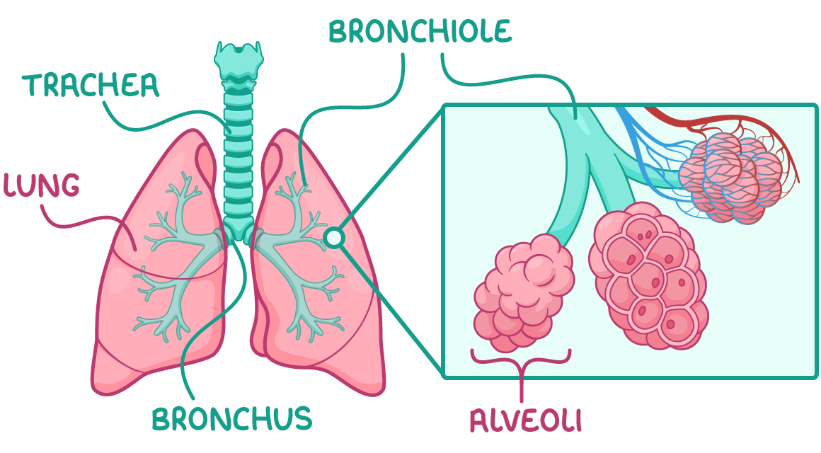
It does this in the following stages:

  1. Air first enters the trachea.
  2. Air travels into the two bronchi, with one bronchus going to each lung.
  3. Air travels into smaller airways called bronchioles.
  4. Air travels into clusters of air sacs called alveoli at the end of the bronchioles.

Ciliated epithelium

A tissue called the ciliated epithelium is located throughout most of the airways.

Diagram showing ciliated epithelium with cilia, goblet cells, and mucus secretion.

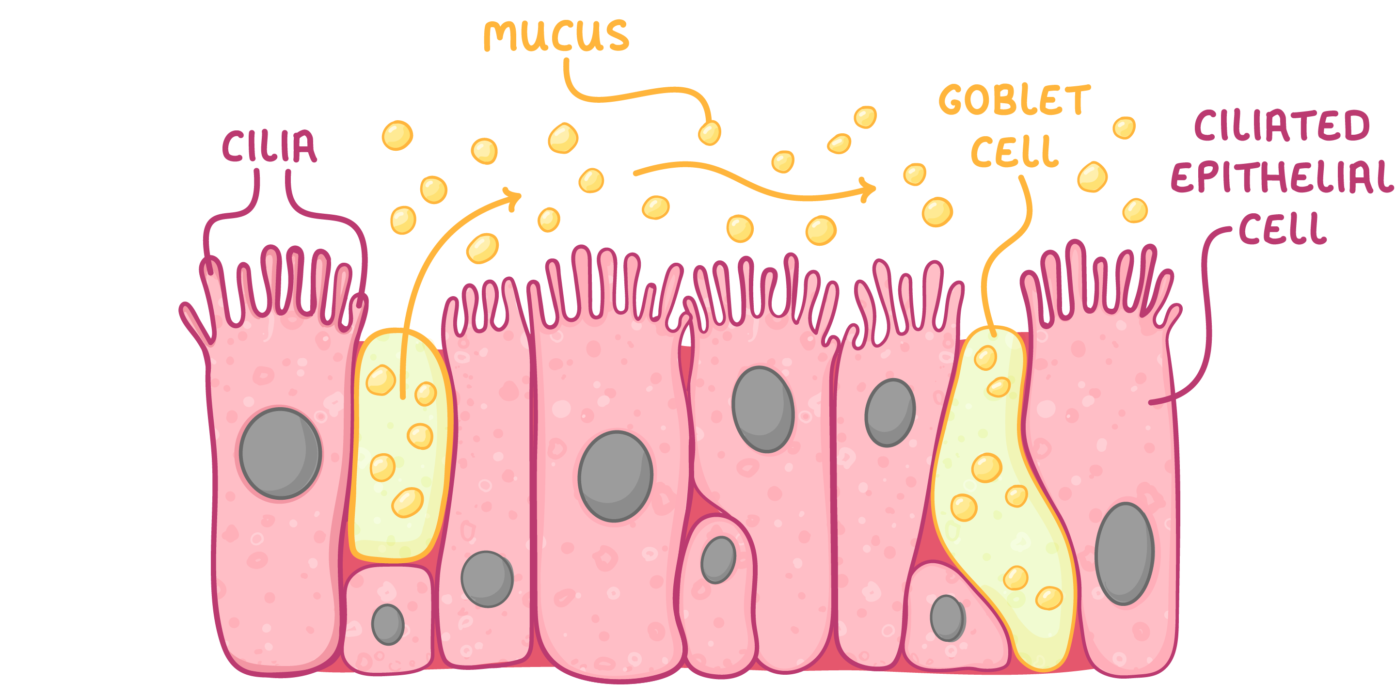
The ciliated epithelium contains mainly goblet cells and ciliated epithelial cells:

  1. Goblet cells - These produce and secrete mucus that traps dust and microbes.
  2. Cilia on ciliated epithelial cells - These waft the mucus upward to the mouth so it can be swallowed.

Structure and adaptations of airways

You need to know about the structure of the trachea, bronchi, and bronchioles, and how these structures help them carry out their functions.

Trachea

The trachea is the large tube that carries air from the throat down to the lungs.


Adaptations:

  1. Rings of cartilage keep the airway open.
  2. Smooth muscle can contract or relax to constrict or dilate the airway and change airflow.
  3. Elastic tissue contains elastic fibres with elastin that allows stretching and recoiling.
  4. Lined with ciliated epithelial cells and goblet cells.

Bronchi

The bronchi are two main branches extending from the trachea that carry air into each lung.


Adaptations:

  1. Reinforced with cartilage to keep the airway open.
  2. Smooth muscle can contract or relax to constrict or dilate the airway and change airflow.
  3. Elastic tissue contains elastic fibres with elastin that allows stretching and recoiling.
  4. Lined with ciliated epithelial cells and goblet cells.

Bronchioles

The bronchioles are smaller airways branching from the bronchi that carry air to the alveoli.


Adaptations:

  1. No cartilage, can change shape.
  2. Smooth muscle can contract or relax to constrict or dilate the airway and change airflow.
  3. Elastic tissue contains elastic fibres with elastin that allows stretching and recoiling.
  4. Simple squamous epithelium (only larger bronchioles have a ciliated epithelium).

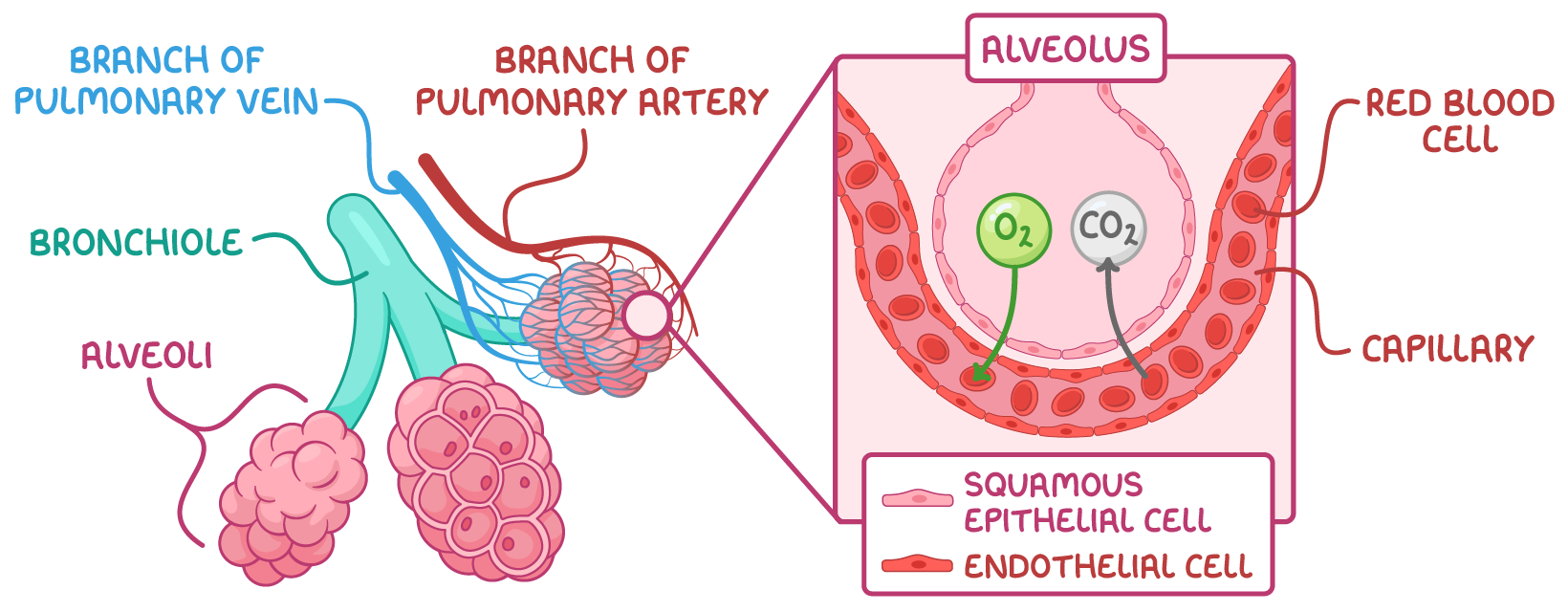
Alveoli and blood vessels

Alveoli are tiny air sacs clustered at the ends of the bronchioles. They are surrounded by a network of capillaries so gases can be exchanged between the air in the alveoli and the blood. 


Gas exchange occurs across the alveolar membrane of alveoli.


This is how the alveoli carry out gas exchange:

  1. Oxygen diffuses from the alveoli into the pulmonary capillaries where it binds to haemoglobin in red blood cells.
  2. Carbon dioxide dissociates from haemoglobin and diffuses from the blood into the alveoli.
Diagram showing alveoli, bronchiole, branches of pulmonary vein and artery, and gas exchange process with oxygen and carbon dioxide in the alveoli.

Adaptations of the alveoli for gas exchange:

  1. Wall consists of one layer of squamous epithelial cells - This allows rapid diffusion.
  2. Large surface area - This increases rate of gas exchange.
  3. Partially permeable - This means that only certain gases can move across the wall.
  4. Surrounded by dense network of capillaries - These bring blood close to air for gas exchange.
  5. Ventilation of air - This maintains steep diffusion gradient.
  6. Elastic fibres - These allow stretching and recoiling.
  7. Collagen fibres - These contain strong collagen that prevents alveoli from bursting and limits overstretching.
  8. Moist inner surface - This allows gases to dissolve, and lung surfactant helps alveoli remain inflated.

Pulmonary blood vessels

The pulmonary blood vessels are those involved in circulation of the lungs.


They include:

  1. The pulmonary artery - This delivers deoxygenated blood from heart to pulmonary capillaries.
  2. The pulmonary vein - This delivers oxygenated blood from capillaries to heart.
  3. The pulmonary capillaries - These are the site of gas exchange between blood and alveoli.

Adaptations of the pulmonary capillaries for gas exchange:

  1. Thin walls (one endothelial cell thick) - This maintains a short diffusion distance.
  2. Red blood cells pressed against capillary walls - This reduces diffusion distance.
  3. Large surface area - This increases diffusion speed.
  4. Movement of blood - This maintains steep diffusion gradient.
  5. Slow blood movement - This allows more time for diffusion.