Structure of Skeletal Muscle
This lesson covers:
- The different types of muscle
- How skeletal muscles act in antagonistic pairs
- The structure of skeletal muscle fibres
- The components within muscle fibres that allow contraction
- The difference between fast-twitch and slow-twitch muscle fibres
The different types of muscle
Muscles, through the contraction of muscle cells, facilitate body movement. While many muscles contract under conscious control, some operate involuntarily, without the need for conscious thought.
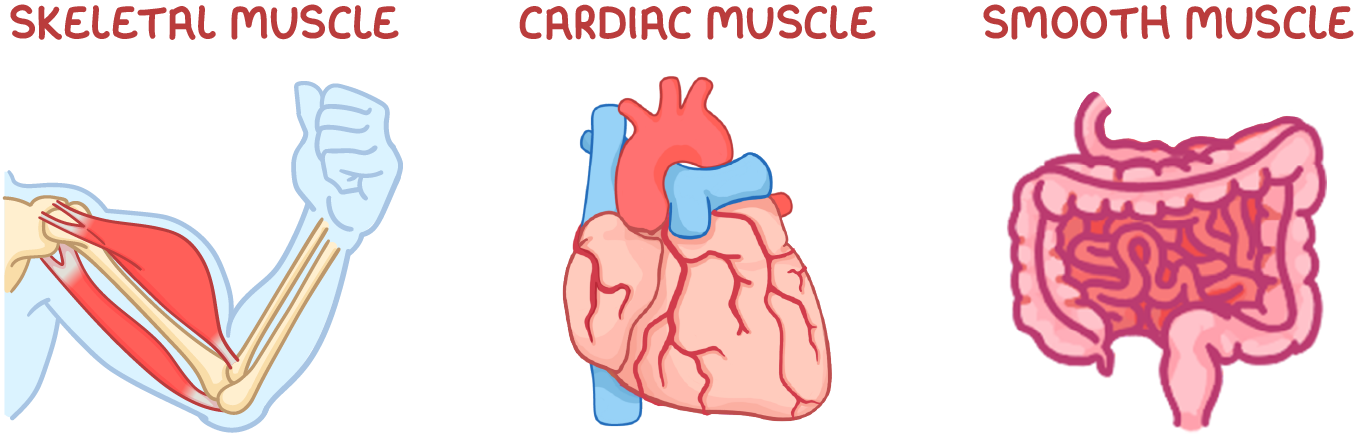
Muscles are categorised into three types:
- Skeletal muscle - This muscle forms the majority of the body's muscles and is attached to bones to voluntarily move parts of the body like the arms or legs.
- Cardiac muscle - This muscle is unique to the heart, and functions to involuntarily circulate blood.
- Smooth muscle - This muscle is located in the walls of hollow organs like blood vessels and the intestines, and typically functions to involuntarily move substances through these organs.
Skeletal muscles act in antagonistic pairs
Skeletal muscles are responsible for moving bones, and they are usually arranged in pairs on opposite sides of a joint, connected to bones by tendons.
These pairs of muscles are known as antagonistic muscles:
- When one muscle contracts, the opposing muscle relaxes.
- The contracting muscle shortens and pulls on its attached tendon.
- This tendon pulls on the bone, causing movement at the joint.
- The relaxing muscle lengthens to allow the movement.
The structure of skeletal muscle fibres
Skeletal muscles comprise numerous bundles of long, cylindrical muscle fibres.
Individual cells are fused to form muscle fibres during development to avoid weakness at the junctions between cells and to increase overall muscle strength.

Key components of a muscle fibre include:
- Sarcolemma - The cell-surface membrane of a muscle fibre.
- Sarcoplasm - The cytoplasm of a muscle fibre.
- Transverse tubules (T tubules) - Extensions of the sarcolemma that transmit electrical signals, to ensure that the entire muscle receives the impulse to contract simultaneously.
- Sarcoplasmic reticulum - A specialised endoplasmic reticulum that is responsible for storing and releasing calcium ions.
- Myofibrils - Subcellular structures designed for contraction.
- Multiple nuclei - Skeletal muscle fibres have many nuclei because several cells merge to form one muscle fibre.
- Mitochondria - These release energy in the form of ATP for muscle contraction.
The structure of myofibrils
Myofibrils, the core units of muscle fibres, contain organised bundles of protein filaments.
These filaments slide past each other to enable muscle contraction.
The structure of a sarcomere
Myofibrils are made up of repeating units called sarcomeres.
Main filaments in a sarcomere:
- Myosin filaments - Thick filaments composed of long rod-shapes with bulbous heads that project to the side.
- Actin filaments - Thin filaments composed of two strands twisted around each other.
Under a microscope, myofibrils exhibit a distinctive banding pattern, which corresponds to the arrangement of myosin and actin within each sarcomere.
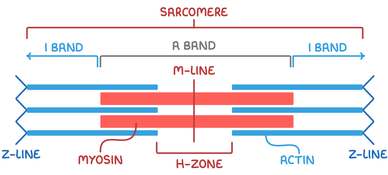
Key sections of the sarcomere:
- A band (dark band) - Area with both myosin and overlapping actin filaments (darker in colour).
- I band (light band) - Area containing only light actin filaments (lighter in colour).
- Z-lines - Mark the boundaries of each sarcomere unit (central line within the I band).
- M-line - The central line of a sarcomere.
- H-zone - Area with only myosin filaments (central lighter region in the A band).
The sliding of actin and myosin filaments within sarcomeres is fundamental to the process of muscle contraction. During muscle contraction, sarcomeres shorten, causing a change in the pattern of light and dark bands.
Slow-twitch and fast-twitch muscle fibres
There are two types of muscle fibres: slow-twitch and fast-twitch muscle fibres. Each of these muscle fibre types have specific adaptations for their roles.
Slow-twitch muscle fibres
Slow-twitch fibres are adapted for endurance work, such as running a marathon.
Their adaptations include:
- Large store of myoglobin - Myoglobin has a higher affinity for oxygen than haemoglobin so carries more oxygen for more efficient aerobic respiration over long time periods.
- Rich blood vessel supply - This provides more oxygen to respiring cells.
- Many mitochondria - This allows more ATP synthesis.
Fast-twitch muscle fibres
Fast-twitch fibres are adapted for intense, short bursts of activity, like weight-lifting.
Their adaptations include:
- More myosin filaments - This increases the ability for muscles to generate a greater force.
- High concentration of glycogen - This can be broken down into glucose to provide more respiratory substrates.
- High concentration of enzymes involved in anaerobic respiration - This allows more anaerobic respiration to occur to release energy quickly.
- Store of phosphocreatine - This can be broken down rapidly to provide phosphates to synthesise ATP during contraction.