Control of Heart Rate
This lesson covers:
- The contraction of cardiac muscle to produce heartbeats
- How the heartbeat is controlled
- The external control of heart rate by the brain and autonomic nervous system
Controlling the heartbeat
Cardiac muscle is myogenic, which means that the contraction of cardiac muscle initiates within the heart itself. The basic rhythm of the heart is maintained by a wave of electrical excitation.
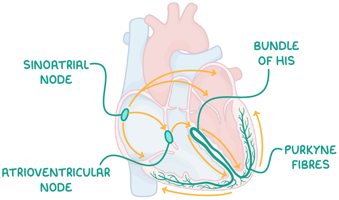
This wave travels through the following structures in this order:
- Sinoatrial node (SAN) - Initiates the heartbeat by stimulating the atria to contract.
- A layer of collagen fibres - Prevents direct electrical flow from atria to ventricles.
- Atrioventricular node (AVN) - Picks up the electrical activity from the SAN and imposes a slight delay.
- Bundle of His - Receives electrical activity from the AVN and conducts the wave of excitation to the apex (base) of the heart.
- Purkyne fibres - These branch off the bundle of His, causing the right and left ventricles to contract from the bottom upwards.
Nervous control of the heart rate
The SAN sets the heart's basic rhythm. The brain and autonomic nervous system can adjust the heart rate based on the body's demands.
The medulla oblongata in the brain stem acts as the control centre. It receives information from receptors in the carotid arteries and aorta.
These receptors are:
- Baroreceptors - Blood pressure receptors.
- Chemoreceptors - Chemical receptors.
The medulla oblongata then regulates the SAN's firing rate to adjust the heart rate.
| Stimulus | Receptors that detect the stimulus | Autonomic pathway | Neurotransmitter used | Effect on SAN | Change in heart rate |
|---|---|---|---|---|---|
| High blood pressure | Baroreceptors | Parasympathetic | Acetylcholine | Decreased electrical activity | Slows down |
| Low blood pressure | Baroreceptors | Sympathetic | Noradrenaline | Increased electrical activity | Speeds up |
| High O2, low CO2, or high pH | Chemoreceptors | Parasympathetic | Acetylcholine | Decreased electrical activity | Slows down |
| Low O2, high CO2, or low pH | Chemoreceptors | Sympathetic | Noradrenaline | Increased electrical activity | Speeds up |
How the autonomic nervous system responds during exercise
This automatic system allows the heart rate to adapt swiftly to the body's changing needs.
For example, during exercise:
- Blood has a higher concentration of carbon dioxide, and so a lower pH.
- Chemoreceptors in the carotid arteries and aorta detect this and increase the frequency of nervous impulses sent to the medulla oblongata.
- The medulla oblongata increases the rate of impulses to the sinoatrial node (SAN) via the sympathetic nervous system.
- Heart rate increases.
- This provides the extra oxygen required for increased respiration in active muscles.
- The increased blood flow also helps remove excess carbon dioxide via the lungs.
- Carbon dioxide concentration reduces.
Hormones, such as adrenaline and noradrenaline, can also influence heart rate. In times of stress, these hormones are released and cause the SAN in the heart to increase the heart rate as part of the 'fight or flight' response.